Is consciousness to be found in quantum processes in microtubules?

Status
Not open for further replies.
What do microtubules "process"? How do they "process" it?
Necessary and sufficient for what? Consciousness? Explain how processing in microtubules produces consciousness.
MT can process (transport) data, MT can store data, MT can integrate and orchestrate data.
Data is the result of sensory reaction producing a stream of data that is transported via microtubules, then compared to memories stored in the brain and "translated" by the brain into a recreation of the sensory data, i.e. an internal image of what the data originally represented. Apparently this evolutionary process resulted in an emergent data sensitivity that was causal to electrochemically elicited emotional experiences.

Anil Seth identifies this "best guessing" by the brain as "controlled hallucinations". I hope you have watched his short but revelatory lecture. He clearly demonstrates how the brain learns and applies knowledge-based successful survival techniques.
What? Explain how a microtubule can "read" or "copy" DNA. Does it do that all by itself?
Yes! MT make exact copies of DNA via the mitotic spindle.

https://www.ted.com/talks/drew_berry_animations_of_unseeable_biology?language=en#t-471663
What? Explain how a microtubule can "process light". What does it "process" light into? How does it do that?
MT in plant leaf perform photosynthesis and distribution of energy.
The Influence of Light on Microtubule Dynamics and Alignment in the Arabidopsis Hypocotyl[W]
Adrian Sambade,a,1 Amitesh Pratap,b,1 Henrik Buschmann,a Richard J. Morris,b and Clive Lloyda,2
Author information Article notes Copyright and License information Disclaimer
This article has been cited by other articles in PMC.

ABSTRACT
Light and dark have antagonistic effects on shoot elongation, but little is known about how these effects are translated into changes of shape. Here we provide genetic evidence that the light/gibberellin–signaling pathway affects the properties of microtubules required to reorient growth.
To follow microtubule dynamics for hours without triggering photomorphogenic inhibition of growth, we used Arabidopsisthaliana
light mutants in the gibberellic acid/DELLA pathway. Particle velocimetry was used to map the mass movement of microtubule plus ends, providing new insight into the way that microtubules switch between orthogonal axes upon the onset of growth.
Longitudinal microtubules are known to signal growth cessation, but we observed that cells also self-organize a strikingly bipolarized longitudinal array before bursts of growth. This gives way to a radial microtubule star that, far from being a random array, seems to be a key transitional step to the transverse array, forecasting the faster elongation that follows. Computational modeling provides mechanistic insight into these transitions. In the faster-growing mutants, the microtubules were found to have faster polymerization rates and to undergo faster reorientations.
This suggests a mechanism in which the light-signaling pathway modifies the dynamics of microtubules and their ability to switch between orthogonal axes.
https://www.ncbi.nlm.nih.gov/pmc/articles/PMC3289555/

Roles of actin cytoskeleton for regulation of chloroplast anchoring
Yuuki Sakai† and Shingo Takagi
Author information Article notes Copyright and License information Disclaimer

ABSTRACT
Chloroplasts are known to maintain specific intracellular distribution patterns under specific environmental conditions, enabling the optimal performance of photosynthesis. To this end, chloroplasts are anchored in the cortical cytoplasm.
In leaf epidermal cells of aquatic monocot Vallisneria, we recently demonstrated that the anchored chloroplasts are rapidly de-anchored upon irradiation with high-intensity blue light and that the process is probably mediated by the blue-light receptor phototropins. Chloroplast de-anchoring is a necessary step rendering the previously anchored chloroplasts mobile to allow their migration.
In this article, based on the results obtained in Vallisneria together with those in other plant species, we briefly discussed possible modes of regulation of chloroplast anchoring and de-anchoring by actin cytoskeleton. The topics include roles of photoreceptor systems, actin-filament-dependent and -independent chloroplast anchoring, and independence of chloroplast de-anchoring from actomyosin and microtubule systems.
Microtubules are motors, as well, now?
Yes! MT are ion powered motors, that drive the flagella for one. This was one of the items in contention during the Kitzmiller v Dover trial. Behe tried to argue that the microtubule motor that drives cilia and flagella was n irreducibly complex organelle, which was debunked by several scientists and caused the judge to rule against the concept of Intelligent Design. That is how I was introduced to one of the many microtubules functions.

Cilia, flagella, and centrioles


cilia1_med_hr.jpeg

Cilia and flagella are projections from the cell. They are made up of microtubules , as shown in this cartoon and are covered by an extension of the plasma membrane. They are motile and designed either to move the cell itself or to move substances over or around the cell. The primary purpose of cilia in mammalian cells is to move fluid, mucous, or cells over their surface. Cilia and flagella have the same internal structure. The major difference is in their length.

http://cytochemistry.net/cell-biology/cilia.htm#
Your unevidence faith in the little things makes them approximately equivalent to elves, I agree.
How about now? If you want more? Just ask.......I love to share......:cool:
 
Last edited:
Weird. That post starts off talking about microtubules, then it moves on to mirror neurons, but nothing in the mirror neuron section mentions microtubules at all. What's special about microtubules in mirror neurons?
That is asking the "hard question" again. The "hard fact" is that mirror neurons do mirror observed behavior in others. Microtubules just do what they do in all neurons, transport sensory data.
 
Last edited:
More on the microtubule motor

Bacterial Flagella: Flagellar Motor

Other versions of this article
Nicolas J Delalez,
First published: 15 August 2014

PDF

Abstract
The bacterial flagellar motor is a complex biological rotary molecular motor, which is situated in the cell envelopes of bacteria. Whereas most biological motors use adenosine triphosphate as their energy source, the rotation of the flagellar motor is driven by a flow of charged ions across the bacterial plasma membrane.
The motor powers the rotation of helical flagellar filaments at speeds of up to several hundred hertz. These rotating filaments act like propellers, pushing the cells through their environment.
The motors are regulated by one of the best-characterised biological signalling pathways, the chemotaxis pathway. This pathway changes the swimming pattern of the bacteria in response to changes in the concentration of external chemicals so that they move into environments, which are optimal for their growth. Other pathways can regulate the flagellar motor and the motor itself can respond to changing conditions by adapting parts of its structure.

Key Concepts:
Many bacteria swim using a small biological rotary motor which is powered by the movement of ions (H+ or Na+) across the plasma membrane.
https://onlinelibrary.wiley.com/doi/abs/10.1002/9780470015902.a0000744.pub4#

The dynein microtubule motor
Stephen MKing

Abstract
Dyneins are large multi-component microtubule-based molecular motors involved in many fundamental cellular processes including vesicular transport, mitosis and ciliary/flagellar beating. In order to achieve useful work, these enzymes must contain motor, cargo-binding and regulatory components. The ATPase and microtubule motor domains are located within the very large dynein heavy chains that form the globular heads and stems of the complex.
Cargo-binding activity involves the intermediate chains and several classes of light chain that associate in a subcomplex at the base of the soluble dynein particle. Regulatory control of dynein motor function is thought to involve the phosphorylation of various components as well as a series of light chain proteins that are directly associated with the heavy chains. These latter polypeptides have a variety of intriguing attributes, including redox-sensitive vicinal dithiols and Ca2+-binding, suggesting that the activity of individual dyneins may be subject to multiple regulatory inputs. Recent molecular, genetic and structural studies have revealed insight into the roles played by these various components and the mechanisms of dynein-based motility.
https://www.sciencedirect.com/science/article/pii/S0167488900000094
 
To illustrate the complete motor assemblies of cilia and flagella;

If you get the "Unavailable" message press "Watch on YouTube"

and for good measure:
 
Last edited:
MT can process (transport) data, MT can store data, MT can integrate and orchestrate data.
In what form is this "data" transported, stored, integrated and orchestrated?

What kind of "data" is it?

Note: saying something like "sensor data" doesn't help here. I understand (to some extent) how sensory organs like eyes and ears produce electrical signals in neurons, for instance. But those do not seem to be processed or transported in any way by microtubules, explicitly, as far as I'm aware. Nerve conduction is happening at a higher level of magnification than the insides of microtubules.

So, what kind of "data" are you referring to.

Data is the result of sensory reaction producing a stream of data that is transported via microtubules...
What is the physical form of the "data stream" you refer to?
Explain how "raw" sensory inputs are translated into this microtubule "data stream".
..., then compared to memories stored in the brain and "translated" by the brain...
"The brain" is a much larger and more complex structure than a microtubule.

What is your evidence that any "data" is "compared to memories" in microtubules? How does that "comparison" occur in the microtubule?

What does a microtubule "translate" and how does it do it? What is the physical mechanism?
... into a recreation of the sensory data, i.e. an internal image of what the data originally represented.
Internal to what?

Is it your claim that human memories are stored specifically in microtubules? Got any evidence for that, at all?
Apparently this evolutionary process resulted in an emergent data sensitivity that was causal to electrochemically elicited emotional experiences.
What evolutionary process? This is your first mention of an evolutionary process.
 
In what form is this "data" transported, stored, integrated and orchestrated?
electrochemical
What kind of "data" is it?
electrochemical
Note: saying something like "sensor data" doesn't help here. I understand (to some extent) how sensory organs like eyes and ears produce electrical signals in neurons, for instance. But those do not seem to be processed or transported in any way by microtubules, explicitly, as far as I'm aware. Nerve conduction is happening at a higher level of magnification than the insides of microtubules.
No, you are making a switch here. Nerve conduction is happening on the outside of the microtubules inside the neurons.

Microtubules in neurons as information carriers - NCBI
Are microtubules inside neurons?

Microtubules in neurons consist of highly dynamic regions as well as stable regions, some of which persist after bouts of severing as short mobile polymers.Dec 11, 2013
https://www.ncbi.nlm.nih.gov/pmc/articles/PMC3979999/

Microtubules – the critical highways of neurons – must stay “clear” for good traffic and neuronal survival
abstract_magda_06_test_export_depuis_applati_et_niveaux.jpg

The excess of tubulin polyglutamylation perturbs transport in primary hippocampal neurons and causes extensive neurodegeneration in mice and humans.
Researchers from Carsten Janke’s group from Institut Curie have recently published two articles of important relevance in the field of Neurobiology.

Neurobiology is the branch of biology, which explores neurons, these tentacle cells also known as nerve cells, which are the primary components of our Nervous System.
A typical neuron1 consists of a cell body, dendrites and an axon2 (see images 1 and 3) 1,4. Most neurons receive signals via the dendrites and send out signals down the axon. However, to allow the spreading of those signals, materials have to be transported to the right place at the right time in the axons.
“Scientists are amazed that microscopic materials can be transported more than several feet along one neuron that goes from the spinal cord to the foot. This is equivalent scale to a person carrying a package walking along the wall of China” 3.
Neuronal cells have to transport mitochondria, vesicles and other materials from one side to the other for their correct functionality. This means from the neuronal cell body to the end of the axon (synaptic terminal) and vice versa. In order to do this, neurons use microtubules as highways along the vast length of their axon.
https://science.institut-curie.org/...clear-for-good-traffic-and-neuronal-survival/
So, what kind of "data" are you referring to
electrochemical.
What is the physical form of the "data stream" you refer to?
electrochemical
Explain how "raw" sensory inputs are translated into this microtubule "data stream".
This is not 'raw" data. It is received and transmitted by the sensors as sorted streams of "patterned" data.
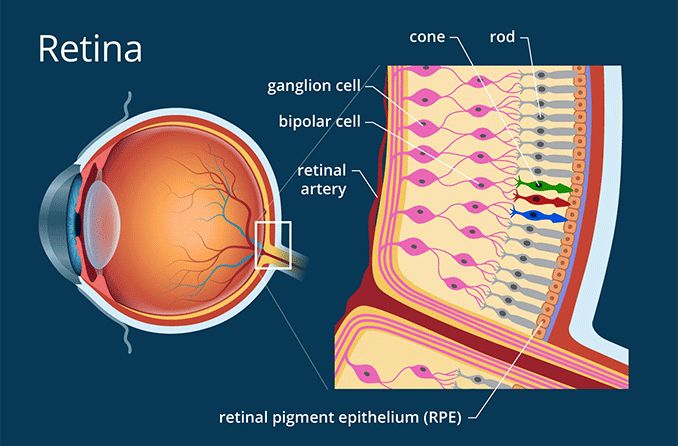
retina-678x446.gif

The first step in the process of vision is the conversion of light into signals that can be interpreted in the brain. This takes place in the retina, which is located in the back of the eye.
https://www.allaboutvision.com/resources/retina.htm
The eye is the first amongst our five senses to be treasured. It is also the smallest and the most complex organ in our body. With its maximum diameter just about 2 cms, generally. The anatomy of the eye includes 2 million moving parts. which is just next to that of the brain. They can distinguish between more than 500 shades of any single colour and see more than 2.7 million colours.
Finally, it hits the sensitive layer of cells called the retina. At the back, it is “encoded” by a light-sensitive membrane called the retina. The cells are called photoreceptors. The retina transforms the image into electrical messages. as electrical impulses to our brain. Turns them into electrical impulses. These impulses are sent to the optic disk on the retina where they get transferred by a further set of electrical impulses along the optic nerve. And sent to the brain to be processed.
https://eyemantra.in/eye-care/anatomy-of-eye/
"The brain" is a much larger and more complex structure than a microtubule.
Is a computer larger than a single processor? The microtubule network in the brain can be counted in the trillions, each with hundreds of dendrites and the synaptic connections in the brain number about 125 trillion.

Stunning details of brain connections revealed
Date: November 17, 2010
Source: Stanford University Medical Center
In particular, the cerebral cortex -- a thin layer of tissue on the brain's surface -- is a thicket of prolifically branching neurons. "In a human, there are more than 125 trillion synapses just in the cerebral cortex alone," said Smith. That's roughly equal to the number of stars in 1,500 Milky Way galaxies, he noted.
https://www.sciencedaily.com/releases/2010/11/101117121803.htm#
What is your evidence that any "data" is "compared to memories" in microtubules? How does that "comparison" occur in the microtubule?
Discovery of quantum vibrations in 'microtubules' inside brain neurons supports controversial theory of consciousness
Date: January 16, 2014
Source: Elsevier
Summary:
A review and update of a controversial 20-year-old theory of consciousness claims that consciousness derives from deeper level, finer scale activities inside brain neurons. The recent discovery of quantum vibrations in "microtubules" inside brain neurons corroborates this theory, according to review authors. They suggest that EEG rhythms (brain waves) also derive from deeper level microtubule vibrations, and that from a practical standpoint, treating brain microtubule vibrations could benefit a host of mental, neurological, and cognitive conditions.
https://www.sciencedaily.com/releases/2014/01/140116085105.htm
What does a microtubule "translate" and how does it do it? What is the physical mechanism?
I don't know how it does that. I do believe that the memories are stored in "pyramidal neural sets of MT" via "phosphorylation".

https://www.ncbi.nlm.nih.gov/pmc/articles/PMC3297561/
Cytoskeletal Signaling: Is Memory Encoded in Microtubule Lattices by CaMKII Phosphorylation?
Travis J. A. Craddock, 1 , * Jack A. Tuszynski, 1 , 2 and Stuart Hameroff 3

Abstract
Memory is attributed to strengthened synaptic connections among particular brain neurons, yet synaptic membrane components are transient, whereas memories can endure. This suggests synaptic information is encoded and ‘hard-wired’ elsewhere, e.g. at molecular levels within the post-synaptic neuron.
In long-term potentiation (LTP), a cellular and molecular model for memory, post-synaptic calcium ion (Ca2+) flux activates the hexagonal Ca2+-calmodulin dependent kinase II (CaMKII), a dodacameric holoenzyme containing 2 hexagonal sets of 6 kinase domains. Each kinase domain can either phosphorylate substrate proteins, or not (i.e. encoding one bit). Thus each set of extended CaMKII kinases can potentially encode synaptic Ca2+
https://www.ncbi.nlm.nih.gov/pmc/articles/PMC3297561/
Internal to what?
Internal in the brain.
Is it your claim that human memories are stored specifically in microtubules? Got any evidence for that, at all?
see above. Check out "pyramidal neurons", which contain clusters of connected MT in pyramidal form.
What evolutionary process? This is your first mention of an evolutionary process.
The evolutionary process that selected greater sensory complexity and ability as a survival advantage (see the eye).

I have posted these results of serious and diligent research several times now, but you keep asking me to explain it, instead of reading the findings of the actual researchers I quote, with links to the actual publication.

Finally, you may want to peruse some of this extensive scientific assessment of the role microtubules play in the brain and indeed throughout the entire body.

https://jonlieffmd.com/blog/are-microtubules-the-brain-of-the-neuron#
 
Last edited:
Note: saying something like "sensor data" doesn't help here. I understand (to some extent) how sensory organs like eyes and ears produce electrical signals in neurons, for instance. But those do not seem to be processed or transported in any way by microtubules, explicitly, as far as I'm aware. Nerve conduction is happening at a higher level of magnification than the insides of microtubules.
I think that was previously addressed ;

Bundles of Brain Microtubules Generate Electrical Oscillations

Large uncompensated charge(10) present in MTs likely plays an important role in electrostatic interactions implicated in MT-macromolecular complexing(11). MTs are thought to generate oscillatory electric fields at expense of elasto-electrical vibrations(12), which may explain our findings that electrically-stimulated MTs behave as biological transistors behaving as sophisticated nonlinear transmission lines, capable of supporting the amplification and axial transfer of electrical signals(13,14,15,16,17,18).
Within the cytoplasm MT-generated variable currents may contribute to the presence and modulation of large intracellular electric fields, which in turn, will help control cell function.
https://www.nature.com/articles/s41598-018-30453-2

Microtubules as Sub-Cellular Memristors
Abstract
Memristors represent the fourth electrical circuit element complementing resistors, capacitors and inductors. Hallmarks of memristive behavior include pinched and frequency-dependent I–V hysteresis loops and most importantly a functional dependence of the magnetic flux passing through an ideal memristor on its electrical charge.
Microtubules (MTs), cylindrical protein polymers composed of tubulin dimers are key components of the cytoskeleton. They have been shown to increase solution’s ionic conductance and re-orient in the presence of electric fields. It has been hypothesized that MTs also possess intrinsic capacitive and inductive properties, leading to transistor-like behavior.
Here, we show a theoretical basis and experimental support for the assertion that MTs under specific circumstances behave consistently with the definition of a memristor. Their biophysical properties lead to pinched hysteretic current–voltage dependence as well a classic dependence of magnetic flux on electric charge. Based on the information about the structure of MTs we provide an estimate of their memristance. We discuss its significance for biology, especially neuroscience, and potential for nanotechnology applications.
Memristors
The term memristor is the contraction of memory and resistor and it was first proposed in 1971 as the fourth element of the electric circuits1. A memristor is defined as a two-terminal passive circuit element that provides a functional relation between electric charge and magnetic flux1,2.
https://www.nature.com/articles/s41598-020-58820-y
 
Last edited:
What evolutionary process? This is your first mention of an evolutionary process.
What humans can do by intent, nature can do by evolution via natural selection, given enough time.
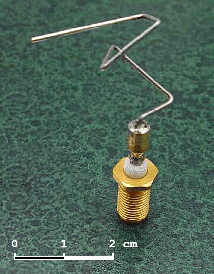
St_5-xband-antenna.jpg

The 2006 NASA ST5 spacecraft antenna. This complicated shape was found by an evolutionary computer design program to create the best radiation pattern. It is known as an evolved antenna.

https://en.wikipedia.org/wiki/Genetic_algorithm

How Does a Fractal Antenna Work?
So, we've seen how fractal shapes repeat themselves over and over and we've seen how antennas can emit energy when electrons are pushed and shoved from one end of an antenna to another, but how do they work together?
Remember, for an antenna it is very important to move the electrons at just the right rate in order to make the antenna work the best. This rate is dependent on the length of the antenna itself. Well, what if we had an antenna that was the same general shape over and over again but at different lengths. This is where the fractal shapes come in. Below you can see a fractal antenna designed to use on an HDTV. See the shape of the wires sticking out? That is a fractal shape known as a Koch Snowflake.
fractal-tv-antenna-515x386.jpg


https://sites.google.com/site/fractalantennas/home/how-does-a-fractal-antenna-work

Note that these shapes often appear in nature. How do they form?

What are Fractals?

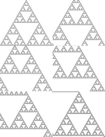
sierpinski-zoom41.gif

They are created by repeating a simple process over and over in an ongoing feedback loop. Driven by recursion, fractals are images of dynamic systems – the pictures of Chaos. Geometrically, they exist in between our familiar dimensions. Fractal patterns are extremely familiar, since nature is full of fractals.

http://xaos-project.github.io/XaoSjs/

https://fractalfoundation.org/resources/what-are-fractals/#
 
The basic functions of a neuron
If you think about the roles of the three classes of neurons, you can make the generalization that all neurons have three basic functions. These are to:
  1. Receive signals (or information).
  2. Integrate incoming signals (to determine whether or not the information should be passed along).
  3. Communicate signals to target cells (other neurons or muscles or glands).
These neuronal functions are reflected in the anatomy of the neuron.

This is an artificially created copper fractal antenna, which can receive and respond to a large range of wavelengths.
2ee683a467ef714df1f948f81b23c9f1.jpg


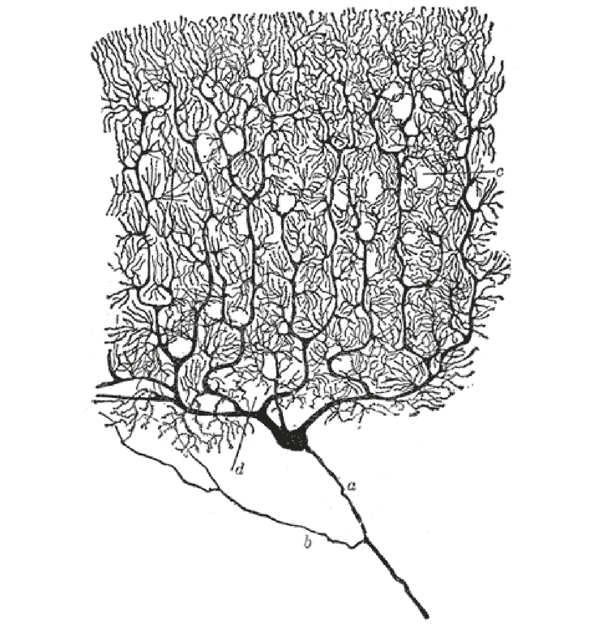
Note that this fractal copper antenna mimics the Purkinje cells in the brain.


e530a086b1674a2d673c562c6d8f3c89619fb4cf.png

For instance, specialized neurons called Purkinje cells are found in a region of the brain known as the cerebellum. Purkinje cells have a highly complex dendritic tree that allows them to receive – and integrate – an enormous number of synaptic inputs, as shown above. Other types of neurons in the cerebellum can also be recognized by their distinctive shapes.
[See a diagram of other cerebellum cell types]

https://www.khanacademy.org/science...m/a/overview-of-neuron-structure-and-function
 
Is it your claim that human memories are stored specifically in microtubules? Got any evidence for that, at all?
Check out "pyramidal neurons", which contain clusters of connected MT in pyramidal form.
In furtherance to the request for evidence, this may clarify:

Local and Global Dynamics of Dendritic Activity in the Pyramidal Neuron

Highlights

1. Discuss the origin of local and global calcium activity in cortical neurons.
2. eview the difficulties and caveats of spatially defining dendritic activity.
3. Reveal evidence in support of both local and global dendritic activity.
4. Illustrate that both local and global dynamics define neural input–output function.

Abstract

There has been increasing interest in the measurement and comparison of activity across compartments of the pyramidal neuron. Dendritic activity can occur both locally, on a single dendritic segment, or globally, involving multiple compartments of the single neuron.
Little is known about how these dendritic dynamics shape and contribute to information processing and behavior. Although it has been difficult to characterize local and global activity in vivo due to the technical challenge of simultaneously recording from the entire dendritic arbor and soma, the rise of calcium imaging has driven the increased feasibility and interest of these experiments.
However, the distinction between local and global activity made by calcium imaging requires careful consideration. In this review we describe local and global activity, discuss the difficulties and caveats of this distinction, and present the evidence of local and global activity in information processing and behavior.

Graphical abstract

1-s2.0-S0306452221003559-ga1.jpg

https://www.sciencedirect.com/science/article/abs/pii/S0306452221003559
A long-range, recurrent neuronal networklinking the emotion regionswith the somatic motor cortex
Yihan Wang1,2and Qian-Quan Sun1,2,3,4,*1Graduate Neuroscience Program, University of Wyoming, Laramie, WY 82071, USA2Department of Zoology and Physiology, University of Wyoming, Laramie, WY 82071, USA3Wyoming Sensory Biology Center of Biomedical Research Excellence, University of Wyoming, Laramie, WY 82071, USA

SUMMARY
Recurrent neural networks (RNNs) are designed to learn sequential patterns in silico, but it is unclear whether and how an RNN forms in the native networks of the mammalian brain. Here, we report an innate RNN, which is formed by the unidirectional connections from three basic units: input units arriving from emotion regions, a hidden unit in the medial prefrontal cortex (mPFC), and output units located at the somatic motor cortex (sMO). Specifically, the neurons from basal lateral amygdala (BLA) and the insular cortex (IC) project to themPFC motor-cortex-projecting (MP) neurons. These MP neurons form a local self-feedback loop and target major projecting neurons of the sMO. Within the sMO, the neurons in the infragranular layers receive stronger input than the neurons in supragranular layers. Finally, we show in vivo evidence that the communications from the emotion regions to the sMO are abolished when MP neurons are chemogenetically silenced
1-s2.0-S2211124721011827-fx1_lrg.jpg


https://reader.elsevier.com/reader/...egion=eu-west-1&originCreation=20211003163531
 
What is your point Write4U , what are you saying ?
I am agreeing with Tegmark and Hameroff that consciousness is an emergent excellence only from inherent physical attributes which all biological forms already possess in various evolutionary stages.

Hameroff and many scientists now confirm that microtubules are biochemical processors of biochemical data.

From plants, to most protists, to all modern Eukaryotic organisms, the microtubule (neural) dynamical information processing network appears to acquire an emergent chemically reactive experience that has evolved into "conscious sentience" at various levels of awareness. And even as in specialized unconscious states such as in the homeostatic "control" of bodily functions in harmony with symbiotic bacterial participation.

Tegmark, and many scientists now confirm a mathematical aspect to all universal expressions and that consciousness is an emergent property of specific neural patterns and processing mechanisms.

Hameroff and Tegmark only disagreed on the potential quantum aspects proposed by Hameroff and Penrose.
I believe that several conceptual conflicts have been resolved in favor of the fundamental ORCH OR concept.

I believe IIT (Giulio Tononi) is a proposal along similar lines.

Integrated information theory

Phi, the symbol used for integrated information
Integrated information theory (IIT) attempts to provide a framework capable of explaining why some physical systems (such as human brains) are conscious,[1] why they feel the particular way they do in particular states (e.g. why our visual field appears extended when we gaze out at the night sky),[2] and what it would take for other physical systems to be conscious (are dogs conscious? what about unborn babies? or computers?).[3]
In principle, once the theory is mature and has been tested extensively in controlled conditions, the IIT framework may be capable of providing a concrete inference about whether any physical system is conscious, to what degree it is conscious, and what particular experience it is having. In IIT, a system's consciousness (what it is like subjectively) is conjectured to be identical to its causal properties (what it is like objectively). Therefore it should be possible to account for the conscious experience of a physical system by unfolding its complete causal powers (see Central identity).[4]
https://en.wikipedia.org/wiki/Integrated_information_theory
 
I have been thinking about your well-meant observation that I am cherry-picking the information that suits my understanding and interpretation.

I totally agree. The reason is that there is only a cherry-tree with low hanging fruit.

When I ask for a counter argument that debunks my perspective, the answer is always that the burden of proof lies with me, and I agree.

The problem is that there are no alternative concepts or theories to be found anywhere. My cherries are the only marketable product available......:rolleyes:
 
This may be of interest;

Note that at 54:00 the artificial construct of measuring brain activity, employs a "coil", a big model of a microtubule.

Now imagine 175 billion of these functioning sensory data processors in a connected network. AI ?

How big is GPT-3?
GPT-3's full version has a capacity of 175 billion machine learning parameters. GPT-3, which was introduced in May 2020, and was in beta testing as of July 2020, is part of a trend in natural language processing (NLP) systems of pre-trained language representations.
https://en.wikipedia.org/wiki/GPT-3

It beat the world champion GO player convincingly, by "learning" to play the game from a lot of recorded games and finally by playing what it learned against itself and spontaneously gaining a masterclass knowledge of the game.
 
Last edited:
This may provide a generalized explanation of Anil Seth's use of the term "controlled hallucination" as a specific evolved survival mechanism that fools the brain into an optical illusion which is beneficial in detecting "camouflaged" predators.

Psychophysics
121px-Psi2.svg.png

Psychophysics quantitatively investigates the relationship between physical stimuli and the sensations and perceptions they produce. Psychophysics has been described as "the scientific study of the relation between stimulus and sensation"[1] or, more completely, as "the analysis of perceptual processes by studying the effect on a subject's experience or behaviour of systematically varying the properties of a stimulus along one or more physical dimensions".[2]
Psychophysics also refers to a general class of methods that can be applied to study a perceptual system. Modern applications rely heavily on threshold measurement,[3] ideal observer analysis, and signal detection theory.[4]
Psychophysics has widespread and important practical applications. For example, in the study of digital signal processing, psychophysics has informed the development of models and methods of lossy compression. These models explain why humans perceive very little loss of signal quality when audio and video signals are formatted using lossy compression.
more.....
https://en.wikipedia.org/wiki/Psychophysics
 
In the Future, We'll Use Glowing Plants as Lamps
Say hello to a legitimately green form of energy.

glowing-plant-with-stem-and-green-leaves-royalty-free-illustration-1633968887.jpg

Nanoparticles injected under the plants' skin can cause them to glow. After charging with light, the plants can glow and recharge indefinitely.
The nanoparticles are coated with silica so that they don't harm the plant, then the scientists "infuse" them through tiny pores in the leaves called stomata (another Greek term, meaning "mouth"). Once inside the plant's cell structure, the particles form a layer that becomes a de facto film. This layer accumulates photons, which are the particles that carry light and other electrical energy.
One of the major improvements over the previous generation of glowing plants comes down to this use of phosphors. The 2017 plant used a combination of the bioluminescent particles luciferin and luciferase, which also help to power the sporadic light that fireflies
produce. These particles were much dimmer, though, and represent a different approach to glowing. Luminescence in general can come from different sources—like the biochemical reaction that happens in fireflies, or the phosphorescence of the 2021 glowing plant.
All of this goes back to the heart of the work: renewable energy.
"Living plants provide an opportunity to rethink the design and fabrication of devices ordinarily produced from plastic and circuit boards and ultimately disposed of as waste," the researchers explain in their paper. "This work establishes methods to transform living plants into photonic substrates for applications in plant-based reflectance devices, signaling, and the augmentation of plant-based lighting."
https://www.popularmechanics.com/science/green-tech/a37887181/glowing-plant-lamp-light-source/

Continued .......
 
continued.....

upload_2021-10-19_2-18-56.png
Figure 5. Changes in Guard Cell Microtubule Structure Number on Stomatal Closing in Darkness and Opening in Light. Numbers of microtubule structures in guard cells and stomatal aperture both decline significantly in darkness following transfer to darkness from the greenhouse, but are restored with subsequent white light treatment (1 h, 100 lmol photons m2 s 1 ). The dark leaf samples were photographed at the end of the dark period then illuminated and used as the 1-h light samples (n = 6). Error bars are standard deviations. Bar = 10 microns

RESULTS
Microtubule Stability and Guard-Cell Function: Microtubule Counts and Order To probe the role of microtubules in guard-cell function, we used confocal microscopy to image microtubule organization in Arabidopsis guard cells expressing GFP-TUA5 (Figure 1).
Stomatal aperture was evaluated by brief visualization using bright-field illumination (see ‘Methods’). Guard cells from open stomata in z-projected images showed more radially arranged microtubule structures (Figure 1B) than guard cells with closed stomata (Figure 1C).
We use the term ‘structure’ to refer both to single microtubules and to co-linear bundles of microtubules that cannot be resolved by optical microscopy. If we count resolved microtubule structures in randomly chosen guard cells from light-grown leaves of greenhouse plants, we find a strong correlation between numbers of these structures and stomatal aperture (R2 = 0.9566, Figure 1A).
On this basis, fewer than 20 microtubule structures were recognizable in z-projections of guard-cell confocal images when no stomatal aperture was visible. Guard cells with open stomata (2 lm) averaged over 50 resolved microtubule structures. When guard cells with open stomata were placed in the dark, stomatal aperture and the number of resolvable microtubule structures both declined by 50% within 30 min (Figure 2). Regression of the curves revealed them to be near linear (R2 = 0.9647 and 0.8667, respectively). Microtubules in surrounding pavement cells were not radially arranged and did not appear to change when stomata closed.
more..........
https://www.researchgate.net/public..._Guard-Cell_Function_in_Vicia_and_Arabidopsis
 
continued.....

View attachment 4479
Figure 5. Changes in Guard Cell Microtubule Structure Number on Stomatal Closing in Darkness and Opening in Light. Numbers of microtubule structures in guard cells and stomatal aperture both decline significantly in darkness following transfer to darkness from the greenhouse, but are restored with subsequent white light treatment (1 h, 100 lmol photons m2 s 1 ). The dark leaf samples were photographed at the end of the dark period then illuminated and used as the 1-h light samples (n = 6). Error bars are standard deviations. Bar = 10 microns

RESULTS
more..........
https://www.researchgate.net/publication/221688241_Microtubules_Are_Essential_for_Guard-Cell_Function_in_Vicia_and_Arabidopsis

Explain to us all , what this all means , Write4U . What are your thoughts .
 
Explain to us all , what this all means , Write4U . What are your thoughts .
I am trying to demonstrate the incredible utility microtubules have in all forms of life.

Clearly, the cytoskeleton is the primary substrate for all dynamic data processing functions in Eukaryotic biology. Plus the fact that brained animals have heavy concentrations of microtubules in brains and neural systems strongly suggests that they are the primary candidate for also being the substrate that yields emergent consciousness.

Most important of all MT is a self-organizing organelle (from just 2 tubulins) and is a "common denominator" in all Eukaryotic biology which must have been a "foundational genetic building block" in the abiogenetic emergence of life itself.
 
Last edited:
Status
Not open for further replies.
Back
Top