Is consciousness to be found in quantum processes in microtubules?

Status
Not open for further replies.
Continuing our journey.

Microtubules as quantum systems?
............
Could DNA sequences code for TQC programs? (as implied in the publication “DNA as Topological Quantum Computer” by M. Pitkanen,, 2010)
Microtubules are reported to have 8 resonance peaks for AC stimulation (kilohertz to 10 megahertz), which appear to correlate with various helical conductance pathways around the geometric microtubule lattice.
The explanation is proposed in terms of current pathways which are identified topological qubits, still why not to speak about braid strands or specify what topological qubit means if one is speaking about TQC?
https://www.academia.edu/17199736/Microtubules_as_Quantum_Systems?email_work_card=abstract-read-more

New results about microtubules as quantum systems
Authors; Matti Pitkanen

Abstract
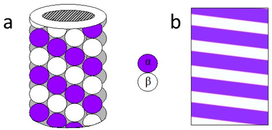
The latest news in quantum biology is the observation by the group led by Anirban Bandyopadhyay about the detection of quantum vibration in microtubule scale - their lengths vary up to 50 nm. If this observation can be replicated, one can speak about a breakthrough in quantum consciousness.
The findings reported in an earlier talk of Bandyopadhyay give support for the general TGD inspired view about topological quantum computation (TQC) and allow for a rather detailed model in the case of microtubules. The idea is that ux tubes form a 2-D coordinate grid consisting of parallel ux tubes in two different directions. Crossing points would be associated with tubulins and the conformational state of tubulin could dene a bit coding whether the braid strands dening coordinate lines are braided or not (swap or not). In this manner any bit pattern at the microtubule denes a particular TQC program.
If also conformations are quantum superposed, one would have "quantum-quantum computation". It however seems that conformation change is an irreversible chemical reaction so this option is not feasible.
The TGD-inspired modification of the proposal in terms of ux tube coordinate grids makes possible TQC architectures with tubulin dimers dening bits dening, in turn, TQC program looks rather natural. Coordinate grids can be xed on the basis of the experimental findings and there are 8 of them. The interpretation is in terms of different resolutions.
The grids for A and B-type lattices are related by 2 twists for the second end of the basic 13-unit for microtubule. An attractive interpretation of the resonance frequencies is in terms of phase transitions between A and B type lattices. If A-type lattices can be generated only in phase transitions induced by AC stimulus at resonance frequencies, one could understand their experimental absence, which is a strong objection against Penrose-Hameroff model.
TGD suggests also a generalization of the very notion of TQC to 2-braid TQC with 2-D string world sheets becoming knotted in 4-D space-time. Now qubits (or their generalizations) could correspond to states of ux tubes dening braid strands as Penrose and Hamero seem to suggest and the emergence of MTs could be seen as an evolutionary leap due to the emergence of a new abstraction level in cognitive processing.
https://dnadecipher.com/index.php/ddj/article/view/6/10
 
And another step

DNA as Topological Quantum Computer: Part I
Author; Matti Pitkanen
Large values of Planck constant makes possible all kinds of quantum computations [47, 41, 48, 50]. What makes topological quantum computation (tqc) [51, 45, 42, 43, 49] so attractive is that the computational operations are very robust and there are hopes that external perturbations do not spoil the quantum coherence in this case. The basic problem is how to create, detect, and control the dark matter with large ~. The natural looking strategy would be to assume that living matter, say a system consisting of DNA and cell membranes, performs tqc and to look for consequences.
There are many questions. How the tqc could be performed? Does tqc hypothesis might allow us to understand the structure of living cell at a deeper level? What does this hypothesis predict about DNA itself? One of the challenges is to fuse the vision about living system as a conscious hologram with the DNA as tqc vision. The experimental findings of Peter Gariaev [57, 59] might provide a breakthrough in this respect. In particular, the very simple experiment in which one irradiates DNA sample using ordinary light in UV-IR range and photographs the scattered light seems to allow an interpretation as providing a photograph of magnetic flux tubes containing dark matter. If this is really the case, then the bottle neck problem of how to make dark matter visible and how to manipulate it would have been ISSN: ISSN: 2159-046X DNA Decipher Journal resolved in principle.
The experiment of Gariaev and collaborators [59] also show that the photographs are obtained only in the presence of DNA sample. This leaves open the question whether the magnetic flux tubes associated with instruments are there in absence of DNA and only made visible by DNA or generated by the presence of DNA.
https://dnadecipher.com/index.php/ddj/article/view/6/10

And microtubules copy DNA during mitosis.
 
Since it is a recent Anil Seth interview, might be of at least slight interest here.
- - - - - - - -

Is consciousness more like chess or the weather?
https://nautil.us/is-consciousness-...6d1-5fe7-4b2b-a7e2-8f63f497ef35.1684463870784

EXCERPTS: I caught up recently with Anil Seth to discuss his work on consciousness, AI, and the worrying intersection of his pair of passions—the possibility of creating conscious AI, machines that not only think but also feel...

[...] Why do you think that consciousness isn’t some kind of complicated algorithm that neurons implement?

This idea, often called functionalism in philosophy, is a really big assumption to make. Some things in the world, when you simulate them, run an algorithm, you actually get the thing. An algorithm that plays chess, let’s say, is actually playing chess. That’s fine. But there are other things for which an algorithmic simulation is just, and always will be, a simulation. Take a computer simulation of a weather system. We can simulate a weather system in as much detail as we like, but nobody would ever expect it to get wet or windy inside a computer simulation of a hurricane, right? It’s just a simulation. The question is: Is consciousness more like chess, or more like the weather?

The common idea that consciousness is just an algorithm that runs on the wetware of the brain assumes that consciousness is more like chess, and less like the weather. There’s very little reason why this should be the case. It stems from this idea we’re saddled with still—that the brain is a kind of computer and the conscious mind is a program running on the computer of the brain.

But the more you look into the brain, the less like a computer it actually appears to be. In a computer you’ve got a sharp distinction between the substrate, the silicon hardware, and the software that runs on it. That’s why computers are useful. You can have the same computer run a billion different programs. But in the brain, it’s not like that at all. There’s no sharp distinction between the mindware and the wetware. Even a single neuron, every time it fires, changes its connection strength. A single neuron tries to sort of persist over time as well. It’s a very complicated object. Then of course there are chemicals swirling around. It’s just not clear to me that consciousness is something that you can abstract away from the stuff that brains and bodies are made out of, and just implemented in the pristine circuits of some other kind of system.
... (MORE - rest of interview)
_
 
Some things in the world, when you simulate them, run an algorithm, you actually get the thing. An algorithm that plays chess, let’s say, is actually playing chess. That’s fine. But there are other things for which an algorithmic simulation is just, and always will be, a simulation. Take a computer simulation of a weather system. We can simulate a weather system in as much detail as we like, but nobody would ever expect it to get wet or windy inside a computer simulation of a hurricane, right? It’s just a simulation.

That's a strange comparison to make. If you open up a chess-playing computer you won't see tiny chess pieces, either. So they are fairly similar; both simulations of a physical system, neither of them has any reality other than that internal simulation.

The common idea that consciousness is just an algorithm that runs on the wetware of the brain assumes that consciousness is more like chess, and less like the weather.

Consciousness is much more like the weather in that we know every single thing we need to know about a chess game to simulate it. You could, for example, take any given position in a chess game, simulate every possible move both the computer and the other person could make in the future, and determine what moves are most likely to win. That takes a lot of computing power but is 100% deterministic.

Weather is a chaotic system. When we simulate it we put in sparse data (temperature at a few locations, past weather, pressure readings, radiosonde readings etc) and then try to simulate it knowing we do not have all the data to start with. So it will be necessarily incomplete - which is why weather forecasts are not 100% accurate. That's similar to our understanding of the brain. We know a few things it's doing and can measure them - but most of what it does is opaque.


But the more you look into the brain, the less like a computer it actually appears to be.
A computer is not at all like a brain. But it is also not at all like a cold front. But it can simulate both to varying degrees of accuracy - which is the issue here.
 
  • Like
Reactions: C C
Just out
Forget AI; Organoid Intelligence May Soon Power Our Computers
William A. Haseltine
Apr 28, 2023,05:27pm EDT
Johns Hopkins University have identified a new form of intelligence: organoid intelligence. A future where computers are powered by lab-grown brain cells may be closer than we could ever have imagined.
What is an organoid? Organoids are three-dimensional tissue cultures commonly derived from human pluripotent stem cells. What looks like a clump of cells can be engineered to function like a human organ, mirroring its key structural and biological characteristics. Under the right laboratory conditions, genetic instructions from donated stem cells allow organoids to self-organize and grow into any type of organ tissue, including the human brain.
Although this may sound like science-fiction, brain organoids have been used to model and study neurodegenerative diseases for nearly a decade. Emerging studies now reveal that these lab grown brain cells may be capable of learning. In fact, a research team from Melbourne recently reported that they trained 800,000 brain cells to perform the computer game, Pong (see video). As this field of research continues to grow, researchers speculate that this so-called “intelligence in a dish” may be able to outcompete artificial intelligence.


https://www.forbes.com/sites/willia...may-soon-power-our-computers/?sh=73fa13995240
 
  • Like
Reactions: C C
Do lab-grown brain cells contain microtubules?

NIH scientists take totally tubular journey through brain cells
Study may advance understanding of how brain cell tubes are modified under normal and disease conditions.

20140613-mircotubule-tagging.jpg

Fantastic Voyage. NIH scientists watched the inside of brain cell tubes, called microtubules, get tagged by a protein called TAT. Tagging is a critical process in the health and development of nerve cells. Roll-Mecak lab, NINDS, Bethesda, MD
In a new study, scientists at the National Institutes of Health took a molecular-level journey into microtubules, the hollow cylinders inside brain cells that act as skeletons and internal highways. They watched how a protein called tubulin acetyltransferase (TAT) labels the inside of microtubules. The results, published in Cell, answer long-standing questions about how TAT tagging works and offer clues as to why it is important for brain health.
Microtubules are constantly tagged by proteins in the cell to designate them for specialized functions, in the same way that roads are labeled for fast or slow traffic or for maintenance. TAT coats specific locations inside the microtubules with a chemical called an acetyl group. How the various labels are added to the cellular microtubule network remains a mystery. Recent findings suggested that problems with tagging microtubules may lead to some forms of cancer and nervous system disorders, including Alzheimer’s disease, and have been linked to a rare blinding disorder and Joubert Syndrome, an uncommon brain development disorder.
“This is the first time anyone has been able to peer inside microtubules and catch TAT in action,” said Antonina Roll-Mecak, Ph.D., an investigator at the NIH’s National Institute of Neurological Disorders and Stroke (NINDS), Bethesda, Maryland, and the leader of the study.
Microtubules are found in all of the body’s cells. They are assembled like building blocks, using a protein called tubulin. Microtubules are constructed first by aligning tubulin building blocks into long strings. Then the strings align themselves side by side to form a sheet. Eventually the sheet grows wide enough that it closes up into a cylinder. TAT then bonds an acetyl group to alpha tubulin, a subunit of the tubulin protein.
Some microtubules are short-lived and can rapidly change lengths by adding or removing tubulin pieces along one end, whereas others remain unchanged for longer times. Recognizing the difference may help cells function properly. For example, cells may send cargo along stable microtubules and avoid ones that are being rebuilt. Cells appear to use a variety of chemical labels to describe the stability of microtubules.
“Our study uncovers how TAT may help cells distinguish between stable microtubules and ones that are under construction,” said Dr. Roll-Mecak. According to Dr. Roll-Mecak, high levels of microtubule tagging are unique to nerve cells and may be the reason that they have complex shapes allowing them to make elaborate connections in the brain.
For decades scientists knew that the insides of long-lived microtubules were often tagged with acetyl groups by TAT. Changes in acetylation may influence the health of nerve cells. Some studies have shown that blocking this form of microtubule tagging leads to nerve defects, brain abnormalities or degeneration of nerve fibers. Since the discovery of microtubule acetylation, scientists have been puzzled about how TAT accesses the inside of the microtubules and how the tagging reaction happens.
more....
https://www.nih.gov/news-events/new...e-totally-tubular-journey-through-brain-cells
 
That's a strange comparison to make. If you open up a chess-playing computer you won't see tiny chess pieces, either. So they are fairly similar; both simulations of a physical system, neither of them has any reality other than that internal simulation.

John Searle said something similar in the past, about simulating the digestion of a pizza not being real digestion. An issue of resolution. A mundane computer simulation isn't reproducing everything down to the level of chemical composition of a pizza and the hydrochloric acid, enzymes, etc -- and beyond to particle substrates below that making the chemical interactions and biological cells ultimately possible. (Though still, not a conventionally tangible pizza one could hold in the hand and literally eat.)

Seth is possibly alluding to something like that when he remarks "Then of course there are chemicals swirling around" -- i.e., neural structure being receptive to their contingent effects of the body's signaling molecules.

But still, the basic aspects of cognitive activity (recognizing and understanding data and reacting to it) can be replicated at a computer's "coarse-grained" level, if not the monkey-wrench thrown in by circulating biochemicals, etc.

A problem with these guys is that they continually keep using the umbrella term "consciousness", rather than narrowing down specifically to what they mean on this and that occasion. Accordingly, I don't see much point in trying to speculatively construe that it's visual, auditory, tactile, etc experiences of consciousness that they might be referring to at times rather than purely information processing "occurring in the dark" (so to speak).

If they continually insist on being that obscure, then they are the source of others potentially misapprehending them (if/when that's even the case).
_
 
Seth is possibly alluding to something like that when he remarks "Then of course there are chemicals swirling around" -- i.e., neural structure being receptive to their contingent effects of the body's signaling molecules.

A problem with these guys is that they continually keep using the umbrella term "consciousness", rather than narrowing down specifically to what they mean on this and that occasion. Accordingly, I don't see much point in trying to speculatively construe that it's visual, auditory, tactile, etc experiences of consciousness that they might be referring to at times rather than purely information processing "occurring in the dark" (so to speak).

I believe that consciousness is no more than an evolved response-ability to sensory and biochemical stimulus.

When a Mimosa or a Venus flytrap reacts to an external disturbance is that an expression of sensory awareness?

At what point does an organism experiencing a biochemical response become conscious of that response?
Is that not just another example of evolutionary refinement of a basic chemical interaction.

Autocatalytic chemical networks at the origin of metabolism
Joana C. Xavier, Wim Hordijk, Stuart Kauffman, Mike Steel, and William F. Martin
Published:11 March 2020

Abstract

Cells are autocatalytic in that they require themselves for reproduction. The origin of the first cells from the elements on the early Earth roughly 4 billion years ago [14] must have been stepwise. The nature of autocatalytic systems as intermediate states in that process is of interest. Autocatalytic sets are simpler than cellular metabolism and produce copies of themselves if growth substrates for food and a source of chemical energy for thermodynamic thrust are provided [57].
In theory, sets of organic molecules should be able to form autocatalytic systems [812], which, if provided with a supply of starting ‘food' molecules, can emerge spontaneously and proliferate via constraints imposed by substrates, catalysts, or thermodynamics [13].
Autocatalytic sets have attracted considerable interest as transitory intermediates between chemical systems and genetically encoded proteins at the origin of life [1317]. Preliminary studies have shown that coenzymes are often required for their own synthesis and are therefore replicators with autocatalytic properties [18]. However, autocatalytic networks have not been identified in non-enzymatic metabolic networks so far, and evidence for their existence during prebiotic evolution is lacking.
https://royalsocietypublishing.org/doi/10.1098/rspb.2019.2377

When the autocatalytic process of a metabolic function goes wrong, this activates another biochemical response of neural discomfort (pain, nausea).

Are the properties of life and consciousness not evolved refinements of sensory awareness and the production of action potentials in response to interaction of information?

Autocatalytic Networks at the Basis of Life’s Origin and Organization
Wim Hordijk1,* and Mike Steel2
Author information Article notes Copyright and License information Disclaimer

Abstract

Life is more than the sum of its constituent molecules. Living systems depend on a particular chemical organization, i.e., the ways in which their constituent molecules interact and cooperate with each other through catalyzed chemical reactions. Several abstract models of minimal life, based on this idea of chemical organization and also in the context of the origin of life, were developed independently in the 1960s and 1970s.
These models include hypercycles, chemotons, autopoietic systems, (M,R)-systems, and autocatalytic sets. We briefly compare these various models, and then focus more specifically on the concept of autocatalytic sets and their mathematical formalization, RAF theory.
We argue that autocatalytic sets are a necessary (although not sufficient) condition for life-like behavior. We then elaborate on the suggestion that simple inorganic molecules like metals and minerals may have been the earliest catalysts in the formation of prebiotic autocatalytic sets, and how RAF theory may also be applied to systems beyond chemistry, such as ecology, economics, and cognition.
Keywords: autocatalytic sets, chemical organization, RAF theory, origin of life
https://www.ncbi.nlm.nih.gov/pmc/articles/PMC6315399/

It seems to me that sensory awareness is a highly evolved system of autocatalytic processes that is being monitored and regulated by the homeostatic control systems. It seems plausible that this process has evolved into a sensitively aware and eventual conscious experience.
 
  • Like
Reactions: C C
Transfer from: http://sciforums.com/threads/do-we-have-freewill-is-it-biblical.165882/page-20#post-3715306

From Whitehead;
"An entity that people commonly think of as a simple concrete object, or that Aristotle would think of as a substance – a human being included – is in this ontology considered to be a composite of indefinitely many occasions of experience."
Could this be what Penrose proposes, in that instead of observation being causal to quantum collapse, quantum collapse is causal to observation (experience)?
The Penrose interpretation is a speculation by Roger Penrose about the relationship between quantum mechanics and general relativity. Penrose proposes that a quantum state remains in superposition until the difference of space-time curvature attains a significant level.[1][2][3]
Penrose's idea is a type of objective collapse theory. For these theories, the wavefunction is a physical wave, which experiences wave function collapse as a physical process, with observers not having any special role. Penrose theorises that the wave function cannot be sustained in superposition beyond a certain energy difference between the quantum states.
Orchestrated objective reduction (Orch OR) is a theory which postulates that consciousness originates at the quantum level inside neurons, rather than the conventional view that it is a product of connections between neurons.
The mechanism is held to be a quantum process called objective reduction that is orchestrated by cellular structures called microtubules. It is proposed that the theory may answer the hard problem of consciousness and provide a mechanism for free will.[1]
The hypothesis was first put forward in the early 1990s by Nobel laureate for physics, Roger Penrose, and anaesthesiologist and psychologist Stuart Hameroff. The hypothesis combines approaches from molecular biology, neuroscience, pharmacology, philosophy, quantum information theory, and quantum gravity.[2][3]
While mainstream theories assert that consciousness emerges as the complexity of the computations performed by cerebral neurons increases,[4][5] Orch OR posits that consciousness is based on non-computable quantum processing performed by qubits formed collectively on cellular microtubules, a process significantly amplified in the neurons.
The qubits are based on oscillating dipoles forming superposed resonance rings in helical pathways throughout lattices of microtubules. The oscillations are either electric, due to charge separation from London forces, or magnetic, due to electron spin—and possibly also due to nuclear spins (that can remain isolated for longer periods) that occur in gigahertz, megahertz and kilohertz frequency ranges.[2][6]
Orchestration refers to the hypothetical process by which connective proteins, such as microtubule-associated proteins (MAPs), influence or orchestrate qubit state reduction by modifying the spacetime-separation of their superimposed states.[7]
The latter is based on Penrose's objective-collapse theory for interpreting quantum mechanics, which postulates the existence of an objective threshold governing the collapse of quantum-states, related to the difference of the spacetime curvature of these states in the universe's fine-scale structure.[8]
Orchestrated objective reduction has been criticized from its inception by mathematicians, philosophers,[9][10][11][12][13] and scientists.[14][15][16] The criticism concentrated on three issues: Penrose's interpretation of Gödel's theorem; Penrose's abductive reasoning linking non-computability to quantum events; and the brain's unsuitability to host the quantum phenomena required by the theory, since it is considered too "warm, wet and noisy" to avoid decoherence.
I believe the warm, wet environment has been resolved.
If correct, the Penrose–Lucas argument leaves the question of the physical basis of non-computable behaviour open. Most physical laws are computable, and thus algorithmic. However, Penrose determined that wave function collapse was a prime candidate for a non-computable process.
In quantum mechanics, particles are treated differently from the objects of classical mechanics. Particles are described by wave functions that evolve according to the Schrödinger equation. Non-stationary wave functions are linear combinations of the eigenstates of the system, a phenomenon described by the superposition principle.
When a quantum system interacts with a classical system—i.e. when an observable is measured—the system appears to collapse to a random eigenstate of that observable from a classical vantage point.
https://en.wikipedia.org/wiki/Orchestrated_objective_reduction
 
Transfer from: http://sciforums.com/threads/do-we-have-freewill-is-it-biblical.165882/page-20#post-3715306

From Whitehead;
Could this be what Penrose proposes, in that instead of observation being causal to quantum collapse, quantum collapse is causal to observation (experience)?

Whitehead was mentioned a couple of times in "The Emperor's New Mind" (along with Russell). But curiously not in a direct relation to consciousness or process philosophy (IEP article).

It is in Chapter 14 of Biophysics of Consciousness: A Foundational Approach (published in 2016) that an association between the two is made. Whitehead is referenced on pages 583, 584, and 519. The excerpt below is from page 519.

Consciousness in the universe: An updated review of the "ORCH OR" theory (PDF)
https://anesth.medicine.arizona.edu...w_of_orch_or_2016_b2237_ch-14_revised-2-3.pdf

Consciousness results from discrete physical events; such events have always existed in the universe as non-cognitive, proto-conscious events, these acting as part of precise physical laws not yet fully understood. Biology evolved a mechanism to orchestrate such events and to couple them to neuronal activity, resulting in meaningful, cognitive, conscious moments and thence also to causal control of behavior. These events are proposed specifically to be moments of quantum state reduction (intrinsic quantum “self-measurement”). Such events need not necessarily to be taken as part of current theories of the laws of the universe, but should ultimately be scientifically describable. This is basically the type of view put forward, in very general terms, by the philosopher Whitehead (1929, 1933) and also fleshed out in a scientific frame-work in the Penrose–Hameroff theory of “orchestrated objective reduction” (“Orch OR”). In the Orch OR theory, these conscious events are terminations of quantum computations in brain microtubules reduced by Diósi–Penrose (DP) “objective reduction” (“OR”), and having experiential qualities. In this view, consciousness is an intrinsic feature of the action of the universe.

I believe the warm, wet environment has been resolved. https://en.wikipedia.org/wiki/Orchestrated_objective_reduction

A quantum biology subdiscipline is still emerging,

(May 15, 2023) Quantum physics proposes a new way to study biology
https://theconversation.com/quantum...ze-our-understanding-of-how-life-works-204995

EXCERPTS: . . . In a complicated, noisy biological system, it is thus expected that most quantum effects will rapidly disappear, washed out in what the physicist Erwin Schrödinger called the “warm, wet environment of the cell.”

[...] Chemists, however, have for a long time begged to differ. Research on basic chemical reactions at room temperature unambiguously shows that processes occurring within biomolecules like proteins and genetic material are the result of quantum effects.

Importantly, such nanoscopic, short-lived quantum effects are consistent with driving some macroscopic physiological processes that biologists have measured in living cells and organisms. Research suggests that quantum effects influence biological functions, including regulating enzyme activity, sensing magnetic fields, cell metabolism and electron transport in biomolecules.

The tantalizing possibility that subtle quantum effects can tweak biological processes presents both an exciting frontier and a challenge to scientists...

_
 
And a long-awaited answer to the question of the electrical properties of microtubules.

Microtubules as One-Dimensional Crystals: Is Crystal-Like Structure the Key to the Information Processing of Living Systems?
Abstract
Each tubulin protein molecule on the cylindrical surface of a microtubule, a fundamental element of the cytoskeleton, acts as a unit cell of a crystal sensor. Electromagnetic sensing enables the 2D surface of microtubule to act as a crystal or a collective electromagnetic signal processing system.
We propose a model in which each tubulin dimer acts as the period of a one-dimensional crystal with effective electrical impedance related to its molecular structure.
Based on the mathematical crystal theory with one-dimensional translational symmetry, we simulated the electrical transport properties of the signal across the microtubule length and compared it to our single microtubule experimental results. The agreement between theory and experiment suggests that one of the most essential components of any Eukaryotic cell acts as a one-dimensional crystal.
Keywords: microtubule; tubulin; crystal; transmission network1.
Introduction
In the past, several theoretical studies were completed by physicists on the vibrational characteristics of bio-living systems, which are currently becoming more attractive research domains in the present. State-of-the-art tools are used to detect vibrational aspects and principle authentication. Traditional theoretical methods are not sufficient to understand the process of biological systems [1,2].
The cooperative interaction between individual components of the developed dynamic system of the biological system changes in a specific fashion [3]. Biological materials show complex vibrational patterns. In other words, solid structures have a crystalline structure and the translational symmetry/spatial periodicity confirms them as an integrated mechanical system, which is well known in solid-state physics or lattice theory.
The lattice, known as the base, can be found by the duplication of building elements [4]. The electrons interact with the atomic lattice with atomic potentials. The electronic bandgap is generated when an electronic state does not exist for a certain energy range [5].
Consequently, a wide range of man-made systems is inspired by the design and organization of such biological systems, which include some of their properties. Whereby we have photonic crystals (crystals in which phonons interact with the periodic variation of elasticity and mass of structure elements) and phononic crystals (crystals in which photons are affected by the variation of the dielectric constant of structure) [6,7].
However, there are many examples of photonic crystals [8] in nature, such as the Chrysinaresplendens beetle in which 120 layers produce color, commonly known as one-dimensional photonic crystals [9]. More, a two-dimensional photonic crystal film is found in the peacock Pavomuticus, which is composed of more than ten-unit cells in thickness, and that layer has a highly reflective nature and provides a saturated color.
The interactive wavelength is determined by the lattice parameters of the layers [10]. Neurofilament and microfilaments are not photonic crystals, although they form the basic features of neurons, they carry out information processing during the cell division process and serve as carriers of vesicles and several components in the biological system.
Crystalline structures are found in various forms in nature. Bressloff PC & Cowan JD [11] present a model of pattern formation in the primary visual cortex (V1) of the brain that considers a crystalline structure.
In order to understand biological systems more broadly, an interdisciplinary approach based on mathematical and physical sciences can make a significant contribution as biological crystal structures are not limited to photonic domains.
The propagation of electromagnetic quanta through a biological lattice leads to a wide range of applications. For example, quantized energy propagation can have applications in wireless electromagnetic fields in medical treatment. The microtubule is the closest candidate related to it.
Microtubules in living cells are organized as networks (where they regularly shrink and grow) and in neurons (where they form highly complex networks with neurons and microfilaments, which generate large-scale communication pathways). To demonstrate the function of cells, microtubules are interconnected through multiple proteins (Figure 1a).
crystals-11-00318-g001-550.jpg

Figure 1. (a) Sketch of a microtubule. (b) Simple drawing of a single photonic crystal layered with 18° tilt.
The outer and inner diameter voids of the microtubule cylinder are approximately 25 nm and 15 nm respectively and they are found in animals from 200 nm to 25 µm [12]. The length of the dimer unit is 8 nm distributed over a Gaussian distribution with a 2 nm standard deviation and 13 protofilaments are present in the cylindrical surface of the microtubule [13].
The protofilaments are tightly bound from the inside and have weak ties between them, forming 2D sheets that are wrapped with a tube through the nucleation process [14]. Each monomer of the lattice of the microtubule has H12- a small C-terminal helix, which emits after a sequence of amino acids that extend from the microtubule surface.
........
Researchers believe that the dissemination of information in neural networks occurs at synaptic junctions, while experimental evidence suggests that microtubules may function as RAM, which demands correction more accurately than at a synaptic junction. It is also noteworthy that a sequence of protein molecules has to undergo a conformational chain in a particular order to open the ion channel.
Therefore, vibration is key to the conventional understanding of membranes and ionic-based information processing. Although the terms of neural oscillations [26,27,28] in brain activity are mostly preferred with electrical impulses through membrane molecules, and the oscillatory term of the brain refers to the rhythm of electrical activity [29,30] in the central nervous system, CNS, and this can form the microtubule from the transmission system of the electromagnetic quanta [31,32].
......
We speculate that brain waves can be thought of as Bloch waves that may provide important mechanisms for consistent large-scale integration between brain regions [17,18,24].
In this work, we study, experimentally and theoretically, electrical transport properties within tubulin-based microtubules (MT). Using the above MTs properties and symmetry we model them as a periodic crystal with a defect and compare the experimental transmission with our model predicted transmission spectrum.
more .... https://www.mdpi.com/2073-4352/11/3/318#

Does this answer the question that the microtubule network is a viable model of data processing in the brain that must consequently be the functional substrate from which consiousness could emerge?
 
Last edited:
And a long-awaited answer to the question of the electrical properties of microtubules. [...] https://www.mdpi.com/2073-4352/11/3/318#

Does this answer the question that the microtubule network is a viable model of data processing in the brain that must consequently be the functional substrate from which consiousness could emerge?

It's a tentative step, but the idea of MTs even being information carriers still seems to be an uphill battle.

Faintly similar to DNA computation, a team of researchers 20 or 30 years from now may have to literally use them in an extracellular context for information processing, to ultimately demonstrate the capacity (if any).

Aside from the dominating neurocentrism preference, there's probably skeptical concern about motivated reasoning in these pursuits. Akin to how, in cosmology, Fred Hoyle and supporters never gave up on the steady state model.

That there may be such a predilection for microtubules having purposes beyond architectural and intracellular transport roles, that it affects and steers setup and interpretation of results in some studies. But by the same token, the pessimism about cognitive bias seeping into such papers potentially also slowing incremental acceptance of positive indications in that direction (if any).
_
 
It's a tentative step, but the idea of MTs even being information carriers still seems to be an uphill battle.
Yes, I can see that in a case of an entirely new concept requiring a mental sea change, but MTs occupy the interior of all Eukaryotic cells, including plants. Why should there be resistance to a better defined knowledge of intracellular , intercellular, and specifically long-range neural communication processes and information sharing?

It is these little analogies that pique my interest.

And then there is the more formal explanation that describes the generalities in more detail. I am spellbound when processing this knowledge of how we acquire knowledge!

Microtubules in neurons as information carriers
Erik W. Dent, PhDcorresponding author1 and Peter W. Baas, PhD3
Author information Copyright and License information Disclaimer
Abstract
Microtubules in neurons consist of highly dynamic regions as well as stable regions, some of which persist after bouts of severing as short mobile polymers. Concentrated at the plus ends of the highly dynamic regions are proteins called +TIPs that can interact with an array of other proteins and structures relevant to the plasticity of the neuron.
It is also provocative to ponder that short mobile microtubules might similarly convey information with them as they transit within the neuron. Thus beyond their known conventional functions in supporting neuronal architecture and organelle transport, microtubules may act as “information carriers” in the neuron.
Microtubules are major architectural elements without which the neuron could not achieve or maintain its exaggerated shape. In addition to serving as structural elements, microtubules are railways along which molecular motor proteins convey cargo.
Microtubule arrays in axons, dendrites, growth cones, and migratory neurons are tightly organized with respect to the intrinsic polarity of the microtubule, which is relevant to both its assembly and transport properties. Vibrant research is being conducted on the mechanisms by which microtubules are organized in different compartments of the neuron, how microtubule dynamics and stability are regulated, and the orchestration of microtubule-based transport of organelles and proteins. While all of this is surely enough to cause one to marvel, we cannot avoid pondering - what other work might microtubules do for neurons?
We are inspired to think about this question by a sizeable body of knowledge about how microtubules and the actin cytoskeleton influence one another. It has long been known that when microtubules are pharmacologically disassembled, the actin cytoskeleton responds, and often dramatically. The engineers have taught us that this response is due, at least in part, to physical principles wherein microtubules bear compressive forces of the contractile actin cytoskeleton, such that the removal of microtubules causes a notable uptick in those forces (Heidemann et al. 1995).
Cell biologists do not disagree, but have argued that the force relationship may have more to do with the balance of forces generated by microtubule-based and actin-based motor proteins (Baas & Ahmad 2001). There is an additional factor, however, which the biochemists might argue is the most important of all. When microtubules are disassembled, they release factors that had been bound to the lattice of the microtubule, and these factors play important roles in signaling pathways that impact the actin cytoskeleton (Wittmann & Waterman-Storer 2001). Such factors may include kinases and small G proteins.
Thus, without minimizing the contribution of physical principles or the importance of motor-driven forces, these latter observations suggest that microtubules are loaded with signaling information. Such a perspective was further buoyed with the discovery of +TIPs (Akhmanova & Steinmetz 2008), as these proteins affiliate with the plus ends of microtubules during bouts of assembly and can interact with a huge variety of other proteins, many of which reside in the cell cortex. Here we ponder whether this theme, of microtubules as information carriers, might be important in a variety of ways in neurons, perhaps every bit as important as the roles microtubules play as architectural elements and railways for organelle transport (Figure 1).

upload_2023-6-2_3-58-7.png
Figure 1
Microtubules as information carriers in the axon and dendrite
Schematic showing microtubules in the axon and dendrite of a stylized neuron. Note the small, stable translocating microtubules (orange) in the axon (left) and the dynamic microtubules invading dendritic spines (right). It is not yet known what proteins the small translocating microtubules in the axon may potentially bind and release (question mark). However, multiple studies have demonstrated dynamic microtubules are capable of polymerizing directly into dendritic spines, concentrating +TIP proteins (yellow stars) during polymerization and releasing them upon depolymerization. See text for details.
[/quote]more .....
https://www.ncbi.nlm.nih.gov/pmc/articles/PMC3979999/
 
Last edited:
A little tidbit about memory processing in neurons (microtubules).

Secret inner workings of cells revealed through self-assembling 'memory' chains
News
By Harry Baker
published January 05, 2023
Researchers genetically altered mouse brain cells to produce physical timelines of their key events in the form of a self-assembling chain of fluorescent proteins.
upload_2023-6-11_0-20-17.pngFluorescent protein chains produced by genetically-altered mouse neurons. (Image credit: University of Michigan)
Researchers have coaxed mouse brain cells into producing self-assembling protein chains that can record information, or "memories," about the hidden processes that take place within the cells. Once fully formed, these biological black boxes can be easily read using a light microscope, which could potentially revolutionize how scientists study cellular processes and the diseases that affect them.
Cells are hubs of constant activity, carrying out the crucial everyday tasks that keep organisms alive. This activity is coordinated by specific "cellular events," such as the expression of certain genes or the triggering of cellular pathways, a series of interactions between molecules in a cell that leads to a certain product or a change in a cell. But understanding exactly how these cellular events play out can be challenging.
RNA or other molecules created during these events inside the cells, scientists have learned how most cellular events work. However, this method provides only a brief snapshot of the event. And, although these snapshots can be stitched together to form a loose picture, scientists likely miss a lot of what is really going on.
https://www.livescience.com/protein-chain-stores-memories-of-cell
 
Another interesting fact about higher brain function.

The microtubule skeleton and the evolution of neuronal complexity in vertebrates
Nataliya I. Trushina , Armen Y. Mulkidjanian and Roland Brandt
From the journal Biological Chemistry
https://doi.org/10.1515/hsz-2019-0149

Abstract
The evolution of a highly developed nervous system is mirrored by the ability of individual neurons to develop increased morphological complexity. As microtubules (MTs) are crucially involved in neuronal development, we tested the hypothesis that the evolution of complexity is driven by an increasing capacity of the MT system for regulated molecular interactions as it may be implemented by a higher number of molecular players and a greater ability of the individual molecules to interact. We performed bioinformatics analysis on different classes of components of the vertebrate neuronal MT cytoskeleton.
Purkinje cells of the cerebellar cortex are an informative example of neurons with a high interconnectivity. They belong to the largest neurons in the human brain and establish the most extensive dendritic arborization in the central nervous system (CNS), which allows for the formation of up to 200 000 synaptic inputs per neuron (Tyrrell and Willshaw, 1992).
The cerebellum is a major part of the hindbrain, which represents an evolutionary old part of the brain and may have first evolved in the last common ancestor of chordates and arthropods between 570 and 555 million years ago (Ghysen, 2003). Thus, the morphological complexity of the dendritic arbor of Purkinje cells can be compared from the brain of simple vertebrates such as lamprey to primates including humans.
Determination of the fractal dimensions of Purkinje cells as a measure for dendritic arborization has been performed for some representative species and revealed an increase over evolutionary time (Figure 1A) consistent with an increase of morphological complexity of neurons during evolution; it should however be noted that a regression analysis with a higher number of species did not reveal a significant increase of the fractal dimensions of Purkinje cells for different water creatures, suggesting that also other variables besides evolutionary time need to be taken into account, at least with respect to the development of the cerebellum (Krauss et al., 1994).

j_hsz-2019-0149_fig_001.jpg

Pyramidal neurons of the cerebral cortex may be another informative example for changes in morphological complexity during evolution. In contrast to the cerebellum, the cerebral cortex has evolved most recently and shows the biggest evolutionary variation (Rakic, 2009). It is the largest site of neuronal integration in the brain and plays a key role in higher cognitive functions such as memory and attention.
During mammalian evolution, the cerebral cortex has established a large relative expansion with an allocation of approximately eight neurons in the cerebral cortex for every neuron allocated to the rest of the brain (Herculano-Houzel et al., 2014).
The most abundant cortical neurons (estimated as 70–80% of the total neural population) represent the pyramidal cells (Defelipe, 2011). A cortical brain region with a well-defined cytoarchitecture and connectivity is the hippocampus, which is involved in the consolidation of information from short-term to long-term memory. Pyramidal neurons in the hippocampal subfield CA1 tend to be the most homogeneous population in the hippocampus with respect to their morphological variation within a species (Ishizuka et al., 1995).
In humans, CA1 pyramidal neurons are considered to have a critical role in autobiographical memory retrieval and for re-experiencing detailed episodic memories (Bartsch et al., 2011).
more .... https://www.degruyter.com/document/doi/10.1515/hsz-2019-0149/html?lang=en
 
Some more pop-market attention on the "publish/lecture or perish" sci and scholarly community that might be of interest here.
- - - - - - - - - -

The hallucination of consciousness: Riccardo Manzotti interviews Anil Seth
https://iai.tv/articles/anil-seth-the-hallucination-of-consciousness-auid-2525?_auid=2020

INTRO: The hard problem of consciousness has puzzled philosophers and neuroscientists alike for decades. Here a philosopher, Riccardo Manzotti, and a neuroscientist, Anil Seth, meet to discuss consciousness, the hallucination of reality, and whether consciousness is inside or outside the brain.

- - - - - - - - - -

Why scientists haven’t cracked consciousness: The science of consciousness still has no theory
https://www.vox.com/future-perfect/...-mind-hard-problem-neuroscience-koch-chalmers

EXCERPTS: In 1998, at the conference of the Association for the Scientific Study of Consciousness (ASSC), the neuroscientist Christof Koch made a bet with the philosopher David Chalmers: by 2023, science would be able to explain how the brain’s tangle of neurons gives rise to the phenomenon we call consciousness. The winner would get a case of wine.

[...] At the 26th ASSC conference this past weekend, 25 years after the initial wager, the results were declared: Koch lost. Despite years of scientific effort — a time during which the science of consciousness shifted from the fringe to a mainstream, reputable, even exciting area of study — we still can’t say how or why the experience of consciousness arises.

[...] the kind of world you’re experiencing when awake is basically the same kind of world you experience in a dream: a hallucination. The difference is that our brains are constantly comparing our waking hallucinations to the sensory input they receive from the outside, fine-tuning the waking dream to keep it in line with what the incoming sensory data suggests is going on beyond our skulls. That’s what the neuroscientist Anil Seth means when he calls consciousness a “controlled hallucination.”

[...] there’s no guarantee that some critical mass of correlations between brain states and feelings can ever tell us how or why consciousness happens. Chalmers suspects that at the conclusion of their renewed bet in 2048, despite all the surrounding progress of insight that’s sure to unfold, the mystery may remain as perplexing as ever...
(MORE - missing details)
_
 
Last edited:
That’s what the neuroscientist Anil Seth means when he calls consciousness a “controlled hallucination.”
He also calls it a "best guess" and that particular scenario begs the question how the brain can detect differences between the sensory data and data from memory. It seems that the decision rests on an emotional response to the results of a electrochemical differential equation that triggers an action potential when the brain actually makes a calculation and can emotionally represent differences in the detail of the data. Does it look threatening or disinterested? What makes it so? Is it young or old, square or round, ... etc?

Based on available information the brain can only make a best guess and then the detailed coding of descriptive features part of the "controlled hallucination" becomes imagined and projected as different holographic patterns in the brain's neural network as expressed reality.

If several people agree with their controlled hallucinations, we call that reality. But there can also be mass uncontrolled hallucinations, and then usually something bad happens.
 
Last edited:
Look what I dug up.

Quantum Resonance in the Synaptic Field

From Chapter 21: Open Ending of the audio version of True Hallucinations by Terence McKenna
Jung wrote, "Though we know from experience that psychic processes are related to material ones, we are not in a position to say in what this relationship consists, or how it is possible at all. Precisely because the psyche and the physical are mutually dependent it has often been conjectured that they may be identical somewhere beyond our present experience." Of what does this relationship consist?
My own hunch, and it is only a hunch, is that an explicitly spatial dimension - of a co-dimension inclusive of our continuum - allows a hologram of other realized forms of organization, far distant, to become visible at certain levels of quantum resonance in the synaptic field. These levels have been damped by selection in favor of more directly relevant lines of information relating to animal survival.
Evolution does not reinforce selectively the ability of an organism to perceive at a distance since such an ability has no selective advantage, unless the information it conveys falls upon the receptors of an organism already sophisicated enough in its use of symbols to abstract concepts for later application.
Thus, these quantum resonances carrying intimations of events at a distance only begin to acquire genetic reinforcement once a species has already achieved sufficient sophistication to be called conscious and mind-possessing.
The use of hallucinogens can be seen as an attempt at medical engineering which amplifies, for inspection by consciousness, the quantum resonance of the other parts of the spatial continuum holographically at hand.
This experience is the vision which the UFOs and psilocybin impart: visions of strange planets, life forms, perspectives and societies, machines, ruins, landscapes. The hierophanies all unfold in a "nunc-stans" that has all space standing in it, like a frozen hologram.
Thus, experimentation with hallucinogens by human beings and the rise in endogenously produced hallucinogens as one advances through the primate phylogeny could both be due to a slow focusing on the phenomenon of imagination. Imagination being the deepening involvement of the species with things beheld but not actually existing in the present at hand.
https://jacobsm.com/deoxy/deoxy.org/h_thunch.htm

Quantum Resonance?
 
Continuing my education in the world of microtubules......
Phragmoplast
From Wikipedia, the free encyclopedia

Phragmoplast and cell plate formation in a plant cell during cytokinesis. Left side: Phragmoplast forms and cell plate starts to assemble in the center of the cell. Towards the right: Phragmoplast enlarges in a donut-shape towards the outside of the cell, leaving behind mature cell plate in the center. The cell plate will transform into the new cell wall once cytokinesis is complete.
The phragmoplast is a plant cell specific structure that forms during late cytokinesis. It serves as a scaffold for cell plate assembly and subsequent formation of a new cell wall separating the two daughter cells. The phragmoplast can only be observed in Phragmoplastophyta, a clade that includes the Coleochaetophyceae, Zygnematophyceae, Mesotaeniaceae, and Embryophyta (land plants). Some algae use another type of microtubule array, a phycoplast, during cytokinesis.[1][2]
Structure[edit]
The phragmoplast is a complex assembly of microtubules (MTs), microfilaments (MFs), and endoplasmic reticulum (ER) elements, that assemble in two opposing sets perpendicular to the plane of the future cell plate during anaphase and telophase. It is initially barrel-shaped and forms from the mitotic spindle between the two daughter nuclei while nuclear envelopes reassemble around them. The cell plate initially forms as a disc between the two halves of the phragmoplast structure. While new cell plate material is added to the edges of the growing plate, the phragmoplast microtubules disappear in the center and regenerate at the edges of the growing cell plate. The two structures grow outwards until they reach the outer wall of the dividing cell.
If a phragmosome was present in the cell, the phragmoplast and cell plate will grow through the space occupied by the phragmosome. They will reach the parent cell wall exactly at the position formerly occupied by the preprophase band.
The microtubules and actin filaments within the phragmoplast serve to guide vesicles with cell wall material to the growing cell plate. Actin filaments are also possibly involved in guiding the phragmoplast to the site of the former preprophase band location at the parent cell wall. While the cell plate is growing, segments of smooth endoplasmic reticulum are trapped within it, later forming the plasmodesmata connecting the two daughter cells. [/quote]
The phragmoplast can be differentiated topographically into two areas, the midline that includes the central plane where some of the plus-ends of both anti-parallel sets of microtubules (MTs) interdigitate (as in the midbody matrix), and the distal regions at both sides of the midline.[3]
more ..... https://en.wikipedia.org/wiki/Phragmoplast

and

Phycoplast
From Wikipedia, the free encyclopedia


Schematic representation of types of cytokinesis in the green algae: 1) Phycoplast formation with cleavage furrow (e.g. Chlamydomonas); 2) Cleavage furrow and persistent telophase spindle (e.g. Klebsormidium); 3) Phycoplast and cell plate formation (e.g. Fritschiella); 4) Persistent telophase spindle/phragmoplast with cell plate formation (e.g. Coleochaete)
The phycoplast is a microtubule structure observed during cytokinesis in members of the Chlorophytina, the largest and most well known subphylum of chlorophyte green algae.
Cytokinesis in green algae occurs via a diverse range of mechanisms, including cleavage furrows in some algae and cell plates in others. Plants (=Chloroplastida) of the clade Phragmoplastophyta (a subgroup of charophytes which includes the land plants, desmids, water silk, stoneworts etc.) use structures called phragmoplasts to organize and guide the growing cell plate. In these plants, the microtubules of the telophase spindle
give rise to the phragmoplast and are oriented perpendicular to the plane of cell division and the forming cell plate. The growth of the cell plate eventually disrupts the telophase spindle (see case 4 in picture).
more.... https://en.wikipedia.org/wiki/Phycoplast


Now visualize what it takes to "govern" the data transfers and destinations to accomplish all these tasks with exquisite fidelity.....
e685505d76272c7843e4305eedcc9f0a8c388ed0.png
...... it boggles the mind.

Yet it also is a powerful argument in support of the notion that there is no better candidate than microtubules as the place of origin of consciousness.

It is entirely conceivable that trillions of synapses are involved in the process of "thinking", i.e. electrochemical data exchanges within the brain. At this level the action potentials for electric and chemical responses (comfort, discomfort) are generated and action potentials are a result of the microtubule data processing system. This is what makes the MT brain a complex biological data processing system that is shared by all Eukaryotic organisms and animal species on earth.
MT systems range from bacteria to plants, from slime mold to humans and whales.
 
Last edited:
Status
Not open for further replies.
Back
Top