Is consciousness to be found in quantum processes in microtubules?

Status
Not open for further replies.
Firstly, a neuron is not a microtutubles. The manner in which you put (neurons) in brackets infers that they are the same thing.
I can't believe it! Totally taken out of context by you!
I am the one arguing that neurons are not the processors except in a most general sense that they form neural networks.
It's microtubules inside the neurons which provide the processing scaffolding. Neurons also have other functions.

NEURON
1280px-Complete_neuron_cell_diagram_en.svg.png


Can you find the hidden microtubules? Do you need another description of the functions of microtubules?
Wanna see how they work?
 
Last edited:
WOW, moved for the second time. It has to be a record.

Free, free at last! Now I am free to explore without limit!!!!!!!!!:rolleyes:
 
And why are you telling me this? This is the specialty of Stuart Hameroff.
You may have missed this but it was presented in Post #1 in this thread.
Watch the videos and links I have provided. I think it is most unfair to ignore the wealth of information I provide and then come to me with profound observations which I presented in the very first post of this thread.
Firstly, Hameroff's speciality is not Alzheimer's. He posited an opinion about microtubules and Alzheimer's. But the disease itself is not his specialty.

Secondly, read the links that I provided.. Particularly the part about the treatments that are targeting the cells and proteins that are released in the brain of those with Alzheimer's, you'd see that they do not work. Reason being that a) no one knows if they cause Alzheimer's and b) if they are a product of the disease itself.

I can't believe it! Totally taken out of context by you!
Here is what you said:

Yes, those are the chemical information, the fuel on which the organisms run, microtubules (neurons) are the processors.

I did not take you out of context. You keep posting like this and it is quite deceptive.

I am the one arguing that neurons are not the processors except in a most general sense that they form neural networks.
It's microtubules inside the neurons which provide the processing scaffolding. Neurons also have other functions.
You have provided no scientific evidence that the microtubules inside neurons "provide the processing scaffolding".

WOW, moved for the second time. It has to be a record.
This thread has only been moved once.

Free, free at last! Now I am free to explore without limit!!!!!!!!!:rolleyes:
Lovely! Now I'll get to be the type of person who tells excited little kids that Santa is not real...
 
I did not take you out of context. You keep posting like this and it is quite deceptive.
No, you're just being dense. My OP specifically says microtubules. Why should I introduce neurons when I previously explained the difference. Your annoying habit to make comments without reading what I present makes you a troll.

Neurons were introduced by others and I specifically made the distinction between neurons and microtubules a longggggggggggggggggggggg time ago.

Get with it, please......o_O
 
You have provided no scientific evidence that the microtubules inside neurons "provide the processing scaffolding"
Yes I have. You"ll just have to find it yourself!

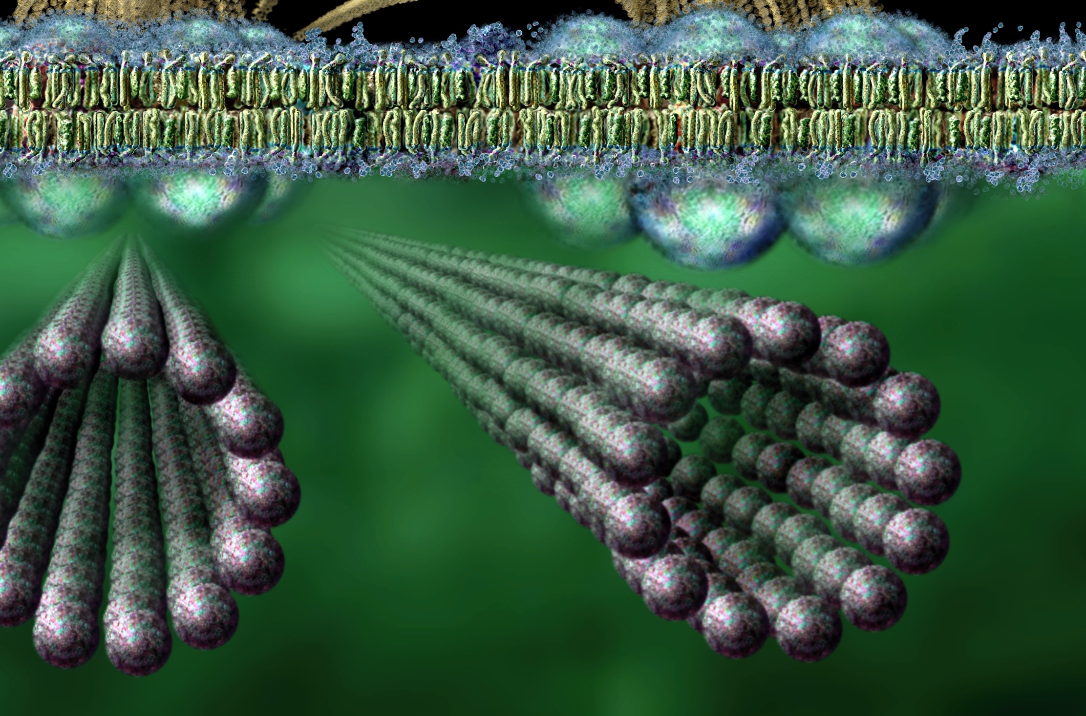
Hint: Drew Berry

click
 
Last edited:
Fot those who wish to continue the inquiry in microtubules.
Microtubules are cylindrical tubes 20-25 nm in diameter, which are composed of linear polymers (protofilaments) of the globular protein tubulin. (right - click to enlarge)
image_filamentous actin microtubules nuclei [] image_filamentous actin & microtubules [] image_microtubules nuclei endothelial tc [] image_filamentous actin microtubules nuclei fibroblast mouse [] image_tubulin microtubules Џ beautiful Flash 8 animation - Inner Life of the Cell, which shows interactions between adhesion-signaling molecules and the cytoskeleton, the scaffolding lattices and conveyor belt mechanisms, and assembly/disassembly of actin and tubulin, and Interpretation: Inner Life of the Cell
https://biologyofcells.blogspot.com/2007/12/microtubules.html
 
And more about "scaffolding".
The Neuron’s “Brain”: The Remarkable Scaffolding Microtubule, The Cells Engineering Language, The Neuron’s Circuit Board[
Microtubule-first-3.jpg


The scaffolding proteins appear to be an engineering language of information flow that immediately responds to mental challenge with changing and evolving physical structures. Are these scaffolding tubules the “brain” inside the neuron? How can mental processes such as attention that alter the neuron’s function instantly affect this structural language of scaffolding molecules to direct rapid changes in dendrites, axons and synapses?
http://jonlieffmd.com/blog/the-neurons-brain-the-remarkable-microtubule-the-cells-engineering-language-the-neurons-circuit-board
 
Discovery of quantum vibrations in 'microtubules' corroborates theory of consciousness
After 20 years of skeptical criticism, "the evidence now clearly supports Orch OR," continue Hameroff and Penrose. "Our new paper updates the evidence, clarifies Orch OR quantum bits, or "qubits," as helical pathways in microtubule lattices, rebuts critics, and reviews 20 testable predictions of Orch OR published in 1998 – of these, six are confirmed and none refuted."
https://phys.org/news/2014-01-discovery-quantum-vibrations-microtubules-corroborates.html


A well deserved bragging after all the knee-jerk reponses by "hasty" critics?
 
Last edited:
No, you're just being dense. My OP specifically says microtubules. Why should I introduce neurons when I previously explained the difference. Your annoying habit to make comments without reading what I present makes you a troll.

Neurons were introduced by others and I specifically made the distinction between neurons and microtubules a longggggggggggggggggggggg time ago.

Get with it, please......o_O
I literally quoted your post back to you.

Yes I have. You"ll just have to find it yourself!

Hint: Drew Berry

click
But you actually have not provided any evidence to support your claims. And the reason for that is simple. Neither have Hameroff and Penrose.

So you keep referring to people who have yet to prove their theories.

Fot those who wish to continue the inquiry in microtubules.
That link does not support any of the claims you have made either by the way..

Discovery of quantum vibrations in 'microtubules' corroborates theory of consciousness

https://phys.org/news/2014-01-discovery-quantum-vibrations-microtubules-corroborates.html


A well deserved bragging after all the knee-jerk reponses by "hasty" critics?
Repetitive.

And no it does not.
 
Continuing on a few items of contention.
Microtubule Network
Throughout the cytoplasm of the eukaryotic cell is a network of microtubles. Cylindrical microtubules are about 25 nm in diameter and lengths range from 200 nm to about 25 micrometers(1). These tubules form part of the cytoskeleton that gives the cell structure and shape. They are also components of cilia and flagella. The microtubule network provides a transportation aid for moving organelles within the cell.
http://hyperphysics.phy-astr.gsu.edu/hbase/Biology/mictub.html

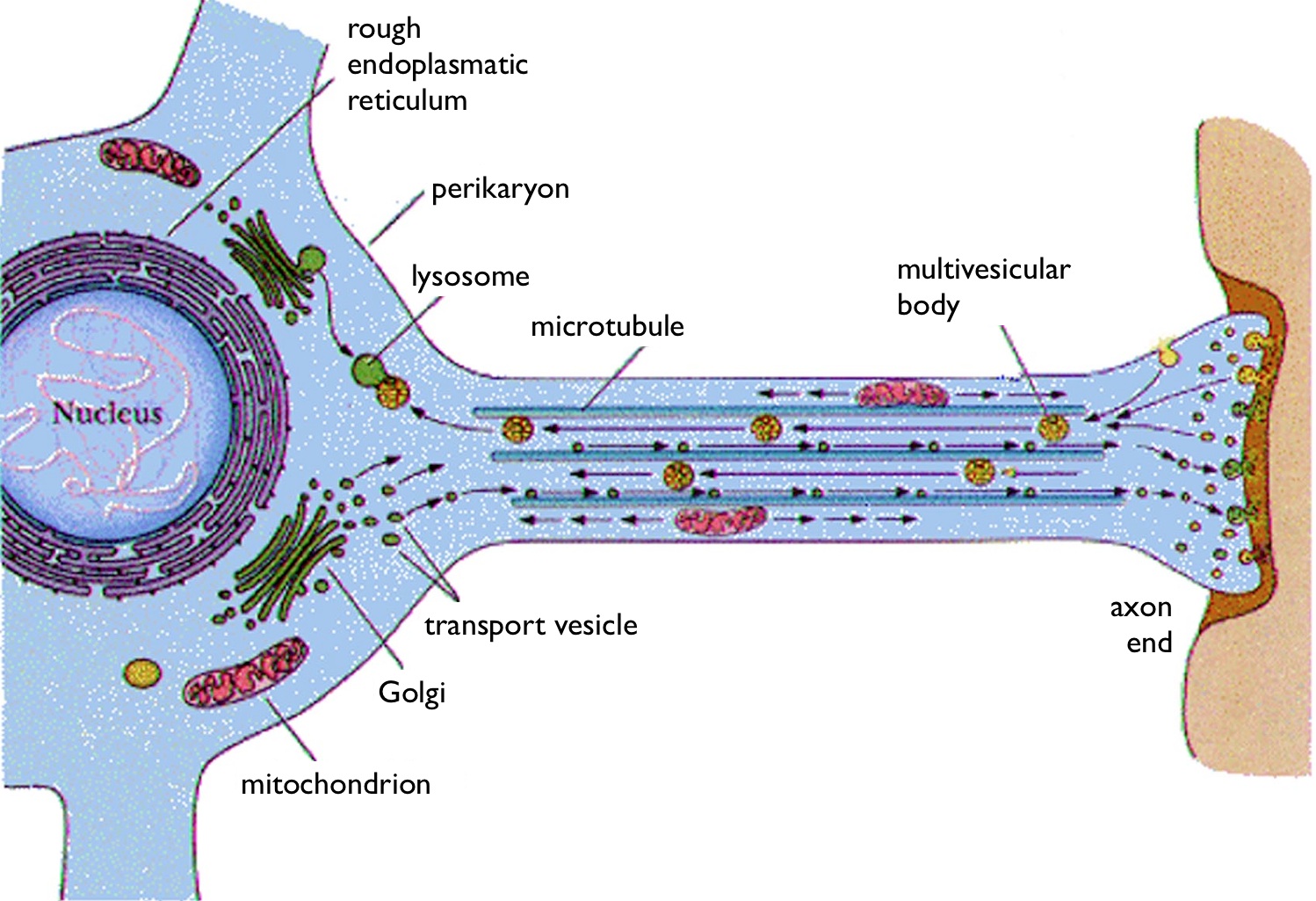
This transportation occurs via microtubules inside all part of neurons and the neural network.
Neural,
1: of, relating to, or affecting a nerve or the nervous system
2: situated in the region of or on the same side of the body as the brain and spinal cord : DORSAL
https://www.merriam-webster.com/dictionary/neural
Neuron,
: a grayish or reddish granular cell that is the fundamental functional unit of nervous tissue transmitting and receiving nerve impulses and having cytoplasmic processes which are highly differentiated frequently as multiple dendrites or usually as solitary axons which conduct impulses to and away from the cell body : NERVE CELL sense
neuron.gif
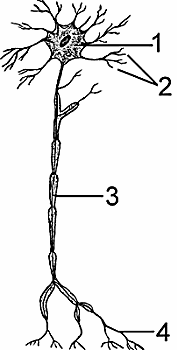
neuron: 1 cell body, 2 dendrite, 3 axon, 4 nerve ending

https://www.merriam-webster.com/dictionary/neuron

Microtubules reside inside the neural cell body, dendrites, axons, nerve endings .
Microtubules are very important in a number of cellular processes. They are involved in maintaining the structure of the cell and, together with microfilaments and intermediate filaments, they form the cytoskeleton. They also make up the internal structure of cilia and flagella. They provide platforms for intracellular transport and are involved in a variety of cellular processes, including the movement of secretoryvesicles, organelles, and intracellular macromolecular assemblies (see entries for dynein and kinesin).[4] They are also involved in cell division (by mitosis and meiosis) and are the major constituents of mitotic spindles, which are used to pull eukaryotic chromosomes apart.
https://en.wikipedia.org/wiki/Microtubule

Microtubules are responsible for the mitosis of neurons themselves.
Microtubules are located in three locations in the cell. They are:
1. Cytoplasmic microtubules which, as the name suggests is located in the cytoplasm.
2. Axonemal microtubules that are located in cilia and flagella
3. Basal bodies, which are structures located at the base of a cilium and contains microtubules.
Generally, microtubules originate from cytoplasmic organelles called centrioles. The region from which microtubules originate is called the microtubule organizing center.

Microtubules and Alzheimers.
upload_2019-10-27_16-59-36.png
https://actualpsychfacts.tumblr.com/image/28554533984

Microtubules are cyclindrical shaped polymers that help maintain and stabilize cell structure. They also have a major role in the transportation of molecules within the cell. They are part of the cytoskeleton, and the cytoskeleton is considered to be an organelle. The microtubules are only a fraction of an organelle under the cytoskeleton.
https://www.answers.com/Q/Are_microtubules_an_organelle
 
Last edited:
Microtubules in plants and direct "orientation"?
Scientists discover important mechanism in plant cells which regulates direction that plant cells grow.
A protein scaffold with the cell, called the microtubule network, dictates the organisation of cellulose in the wall by forming tracks which guide its placement.
Previous work found that microtubules are organised into aligned configurations, with poorly aligned microtubules being cut away by an enzyme called katanin.
https://phys.org/news/2013-09-scientists-important-mechanism-cells.html

Cell Biology: Marine Yeasts Deepen the Sea of Diversity
Effect of microtubules and cell shape on Cdc42 in Schizosaccharomyces pombe. (a) Microtubule bundles align longitudinally within the cell, with plus ends pointing toward cell tips and minus ends in the cell middle. (b) Tip factors (Tea1, Tea4) are delivered to cell ends and bring binding partners, including protein phosphatase 1 (PP1), which promotes localization of the GDP/GTP exchange factor (GEF) Gef1 and exclusion of the GTPase-activating protein (GAP) Rga4 from cell ends. The spatial separation of GEF and GAP promotes Cdc42 activation at cell ends and inactivation at cell sides. (c) Cdc42 is further concentrated by the action of a positive feedback loop similar to that in Saccharomyces cerevisiae, with Scd1 = Cdc24 and Scd2 = Bem1.
https://www.ncbi.nlm.nih.gov/pmc/articles/PMC5944360/figure/F5/

If microtubules are responsible for "orientation", might that suggest potential for "sensitivity to fields", such as EM fields, magnetic fields, gravitational fields?

Is it then a big leap to propose that "navigation by migrating birds might be "guided" by microtubular orientation sensitivity? Or phototropism in plants?
pho·tot·ro·pism,
noun,
1.the orientation of a plant or other organism in response to light, either toward the source of light (positive phototropism) or away from it (negative phototropism).
 
Last edited:
Further confirmation of microtubule catastrophe in Alzheimer patients.

FROM THE MARCH 2015 ISSUE
Cracking the Alzheimer's Code
When Alzheimer examined thin slices of her brain under a microscope, he noticed that nestled right next to the labyrinth circuitry of healthy nerve cells were small clumps of hard, barnacle-like bundles of proteins called amyloid plaques and that many of the fibers extending from the ends of the nerve cells -- (microtubules) -- different proteins called tau — were thickened and tangled. This aberrant brain circuitry — the amyloid plaques and tau tangles — became the twin hallmarks of the disease that bears his name.
(bracketed highlight mine)

neurons-diagram2.jpg

http://discovermagazine.com/2015/march/16-cracking-the-alzheimers-code
 
Alzheimers continued;

Hypothesis: microtubules, a key to Alzheimer disease
Alzheimer disease (AD) is a clinicopathologic syndrome of unknown etiology with numerous abnormalities in neuronal and nonneuronal cells. A review of the literature suggests that a common basic intracellular defect may underlie many of the reported abnormalities.
We hypothesize impairment of the microtubule (MT) system as one explanation for the pathogenesis of AD. Evidence in support of the hypothesis includes the following: MTs are ubiquitous and vital cell components, unequally distributed, with the highest concentration in the brain; various abnormalities, including the key neuropathologic lesions, can be explained by impairments of the MT system; and experiments utilizing pharmacologic agents known to disrupt MTs have reproduced certain abnormalities observed in AD. The hypothesis provides a framework for systematic investigations of MTs at the cellular and molecular levels as well as the basis for in vivo diagnostic tests for AD.
https://www.ncbi.nlm.nih.gov/pmc/articles/PMC298233/
 
Further confirmation of microtubule catastrophe in Alzheimer patients.

FROM THE MARCH 2015 ISSUE
Cracking the Alzheimer's Code
When Alzheimer examined thin slices of her brain under a microscope, he noticed that nestled right next to the labyrinth circuitry of healthy nerve cells were small clumps of hard, barnacle-like bundles of proteins called amyloid plaques and that many of the fibers extending from the ends of the nerve cells -- (microtubules) -- different proteins called tau — were thickened and tangled. This aberrant brain circuitry — the amyloid plaques and tau tangles — became the twin hallmarks of the disease that bears his name.

(bracketed highlight mine)

http://discovermagazine.com/2015/march/16-cracking-the-alzheimers-code

The actual quote:

When Alzheimer examined thin slices of her brain under a microscope, he noticed that nestled right next to the labyrinth circuitry of healthy nerve cells were small clumps of hard, barnacle-like bundles of proteins called amyloid plaques and that many of the fibers extending from the ends of the nerve cells — different proteins called tau — were thickened and tangled. This aberrant brain circuitry — the amyloid plaques and tau tangles — became the twin hallmarks of the disease that bears his name.

The fibers that extended from the end of the nerve cells that were thickened and tangled were not microtubules. What he saw were the "different proteins called tau".. And we know this because when you get to the later parts of the paragraph, it actually says "tau tangles".

Your insertion of "microtubules" into that paragraph altered the actual meaning and context of the entire piece you quoted.

Would you mind explaining why you openly lied and attempted to mislead in such a fashion?
 
The fibers that extended from the end of the nerve cells that were thickened and tangled were not microtubules. What he saw were the "different proteins called tau".. And we know this because when you get to the later parts of the paragraph, it actually says "tau tangles".
The "tau tangles" are not the ends of the microtubules. They are shown in blue after breaking off from the microtubule which is extending from the axon, showing the catastrophe in process, with the "tau particles" falling off the microtubule. The illustration identifies all the parts of the process, by NAME!!!!!!!
Put on your reading glasses.
Your insertion of "microtubules" into that paragraph altered the actual meaning and context of the entire piece you quoted.
No, no, it clarifies any confusion in identification. I did that for your benefit, but as with the problems you had in separating microtubules from neurons, you're misinterpreting the use of the term "nerve cells". I merely made a clarification. You're not paying attention.

It is the microtubule that is disintegrating, not the nerve cell itself. The tau proteins fall off the microtubule, not the nerve cell, and form the tau tangles at the very bottom of the illustration. There is one "tangle" at the bottom left and another "tangle" at the bottom right part of the illustration.

The picture at upper left is a healthy neuron and one of its microtubules with tau proteins assisting in growth of the microtubule.
Note: upper left says "healthy neuron", the lower right picture says "diseased neuron" at top and "disintegrating microtubule" at bottom.

It also shows the discoloration of the stressed neural cell itself, producing the plaque, perhaps?

Pay closer attention. You'll won't look so confused as you appear now.
 
Last edited:
@ Gawdzilla Sama,

Apparently the microtubules also play a part in the formation and location of blood-vessels.

Being Able To Build Human Blood Vessels As Organoids From Stem Cells Is A Game Changer
The more that is learned about how microfluidic processes control or contribute to cellular change, the sooner science will be able to design a cost-effective medical treatment based on that information. New research on microtubules and blood-vessel organoids augments this.
Recent findings by a team of engineering and medical scientists at Stanford University shed new light on how cell components move around and self-renew. Part of the study’s focus was on the link between microtubules and self-organization.
Microtubules, hollow tubes about 25 nm in diameter, are critical for maintaining cell shape and movements. The study analyzed what role microtubules have in maintenance, and found that microtubules are continuously losing and gaining molecules. Understanding the mechanics of how microtubules move around and contribute to regeneration helps to light affordable paths to wound healing in humans and animals.
https://ufluidix.com/circle/being-a...-organoids-from-stem-cells-is-a-game-changer/

It's uncanny, how microtubules keep turning up in biological research.
 
Last edited:
Another source for importance of microtubules in vascular health.

Anti-vascular actions of microtubule-binding drugs
More recent studies have shown that most MBDs also have anti-angiogenic or vascular-disrupting activities or both; in this review these will be referred to collectively as anti-vascular effects (Table 1). Targeting of the tumor vasculature as a therapeutic approach has a compelling theoretical rationale, is strongly supported by pre-clinical studies, and has been validated by clinical trials (34).
There is now data to suggest that MBDs would be a particularly useful class of drugs for this purpose, most notably, evidence that MBDs have multiple direct actions on endothelial cells (discussed in detail below), and that they can produce a much greater reduction in blood flow in tumors than in normal tissues (35). A critical unanswered question is whether the anti-vascular actions of the MBDs contribute to their clinical efficacy, relative to their direct cytotoxic actions against tumor cells. In this regard, there is data from mouse models to suggest that the anti-vascular actions of the MBDs are therapeutically important (36)
https://www.ncbi.nlm.nih.gov/pmc/articles/PMC2745203/
 
The "tau tangles" are not the ends of the microtubules. They are shown in blue after breaking off from the microtubule which is extending from the axon, showing the catastrophe in process, with the "tau particles" falling off the microtubule. The illustration identifies all the parts of the process, by NAME!!!!!!!
Put on your reading glasses.
That's nice.

But I was not addressing the image.

I was addressing the manner in which you altered a quote to make it appear as though it was talking about microtubules when it clearly originally was not.

You inserted "(microtubules)" in bold, altered that sentence, and the manner in which you did it gave the appearance that it was talking about microtubules. It was not.

The manner in which you post and reply is deliberately deceptive and dishonest.

Your first comment in that post alluded directly to microtubules:

Further confirmation of microtubule catastrophe in Alzheimer patients.

You then deliberately altered that quote by inserting "(microtubules)" after these words from the quote "that many of the fibers extending from the ends of the nerve cells "...

The quote I am addressing and the manner in which you deliberately misrepresented that quote by altering it to suit your narrative, is what is deceptive.

Not to mention the fact that you gave the impression that the article was about microtubules, or "microtubule catastrophe", when it clearly was not.. Just as your use of "microtubules catastrophe" in the context that you are using that term is deceptive.

Let me be clear, Write4U..

You altered a quote from an article and made it appear to be something else altogether..

It is blatantly dishonest and deceptive.

No, no, it clarifies any confusion in identification. I did that for your benefit, but as with the problems you had in separating microtubules from neurons, you're misinterpreting the use of the term "nerve cells". I merely made a clarification. You're not paying attention.

It is the microtubule that is disintegrating, not the nerve cell itself. The tau proteins fall off the microtubule, not the nerve cell, and form the tau tangles at the very bottom of the illustration. There is one "tangle" at the bottom left and another "tangle" at the bottom right part of the illustration.

The picture at upper left is a healthy neuron and one of its microtubules with tau proteins assisting in growth of the microtubule.
Note: upper left says "healthy neuron", the lower right picture says "diseased neuron" at top and "disintegrating microtubule" at bottom.

It also shows the discoloration of the stressed neural cell itself, producing the plaque, perhaps?

Pay closer attention. You'll won't look so confused as you appear now.
Stop lying. I was addressing the quote. And it was quite clear that I was - given I posted the quote.. In its original format and the altered version that you posted.

Secondly, the image mentions microtubules because it is a part of the cell. You posted an altered quote, with a sentence about "microtubules catastrophe" and inserted "microtubules" in that quote to alter its meaning and context entirely.

This is basically what it comes down to.

I get it, you are all up about microtubules.

But when you resort to lying and being as dishonest as you have been, you simply come across as a zealot. And if you do it again, I'll issue you with an infraction and shut the thread down.

It's as simple as that.

If you cannot support your claims or argument without outright lying and that level of dishonesty, I'd suggest you refrain from making such claims.
 
Status
Not open for further replies.
Back
Top